


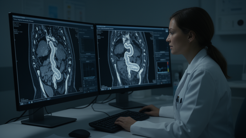
Two of the most widely used imaging tests for Crohn’s disease cannot be relied upon to give matching answers when individual measurements are compared, according to a large multicentre agreement study published in European Radiology Abdomen. MR enterography, an MRI scan of the bowel, and intestinal ultrasound are both recommended as first-line investigations for diagnosing Crohn’s disease and monitoring treatment, yet when researchers from the METRIC trial compared the two techniques side by side in the same patients, they found clinically important disagreement for mural thickness and disease length, two of the most commonly reported parameters. Only when readers made a holistic judgement of disease severity in a bowel segment did the two modalities agree reliably, with 95 percent concordance in patients whose disease was visible on both scans.
Between December 2013 and September 2016, 284 patients aged 16 years and older, with either newly diagnosed Crohn’s disease or a suspected relapse, were recruited from eight UK teaching and general hospitals. Each patient underwent both MR enterography and intestinal ultrasound within four weeks of one another, and independent practitioners interpreted each scan blind to the results of the other modality and to the reference standard. The cohort had a median age of 36 years, and 154 of the 284 patients, or 54 percent, were female. Of the participants, 133 were new diagnoses and 151 were suspected relapses, and the reference standard judged disease to be in the terminal ileum in 217 of the 284 patients, the location of most cases in this study.
The imaging was read by 28 study practitioners, 27 of them gastrointestinal radiologists with a median of nine years of experience, and one sonographer with twenty years of experience. For each affected bowel segment, the readers recorded a predefined set of disease activity parameters: ten for MR enterography and fourteen for intestinal ultrasound. The researchers mapped these parameters between the modalities, since some signs, such as bowel wall thickness, are directly comparable, while others, such as mural oedema seen on MRI, have only approximate equivalents on ultrasound, reflecting the different physical principles of the two techniques. Readers also documented disease length, the presence of obstruction, defined as upstream bowel dilation, and a global segmental disease severity classification of none, mild or advanced. A multidisciplinary consensus panel, including at least one gastroenterologist and one radiologist from the recruitment site plus an external radiologist, served as the reference standard, reviewing all available clinical and imaging data up to six months after recruitment, including colonoscopy and histology when available.
One methodological feature sets this analysis apart from most previous comparisons. Earlier studies typically restricted their analysis to patients in whom both modalities detected disease, which the authors argue introduces bias, because disease missed by one or both tests may be intrinsically different from disease that is found. To address this, the team analysed the full cohort of 284 patients using imputation, assuming that any disease missed by a modality was inactive, with mural thickness set to one millimetre and disease length to zero centimetres. This produces a theoretical worst-case scenario for agreement, because it incorporates all perceptual and technical errors. The researchers then performed a separate complete case analysis limited to the 158 patients in whom both MR enterography and intestinal ultrasound had identified terminal ileal disease and activity parameters had been recorded for both scans.
The numbers reveal a striking gap between the two approaches. In the full cohort, mean mural thickness was 4.9 millimetres on MR enterography and 4.0 millimetres on ultrasound, a mean difference of 0.9 millimetres, but the 95 percent limits of agreement stretched from minus 4.8 to 6.7 millimetres. In the 155 patients with paired data from both modalities, mean thickness was 7.1 millimetres on MRI and 5.9 millimetres on ultrasound, with a mean difference of 1.2 millimetres and limits of agreement of minus 4.0 to 6.3 millimetres. For disease length, the discrepancy was even wider: across all patients, mean length was 9.1 centimetres on MRI and 8.7 centimetres on ultrasound, with 95 percent limits of agreement spanning minus 22.8 to 23.6 centimetres.
The clinical consequences depend on where the measurements fall. The researchers divided mural thickness into threshold categories, using the consensus threshold of three millimetres for normal mural thickness as a reference point. When mean mural thickness was between two and four millimetres, only 24 percent of measurements in the full cohort were within two millimetres of each other, although this rose to 85 percent in the complete case analysis. At thicknesses above four millimetres, 61 percent of full cohort measurements and 66 percent of complete case measurements were within two millimetres. The authors note that disagreement at greater thicknesses may matter less, since a reading of seven versus nine millimetres would suggest active disease either way, but small differences at lower thicknesses could affect the assessment of early treatment response, the calculation of disease activity scores, and even the classification of disease as active or inactive, potentially leading to incorrect treatment decisions in a treat-to-target approach.
Agreement for disease length followed a similar pattern, being better for shorter segments. At mean lengths of five centimetres or less, measurements were within five centimetres of each other in 92 percent of full cohort cases and 100 percent of complete cases, with limits of agreement of minus 5.7 to 6.3 centimetres and minus 3.2 to 2.4 centimetres respectively. At lengths between five and ten centimetres, agreement fell to 69 percent and 82 percent, and for lengths above ten centimetres, only 41 percent of full cohort and 43 percent of complete case measurements were within five centimetres. This pattern echoes an earlier prospective study of 120 small bowel Crohn’s disease patients, which found that intestinal ultrasound underestimated disease length by an average of 30 percent and appeared significantly less accurate for this measurement.
For the categorical activity parameters, one sign stood out. Increased mural T2 signal on MRI, which corresponds to mural oedema, agreed with submucosal layer thickening on ultrasound in 91 percent of cases, 141 of 155. By contrast, other potential ultrasound equivalents of increased mural T2 signal, such as submucosal clarity, agreed in only 50 to 58 percent of cases, and increased perimural T2 signal showed limited agreement of 32 to 48 percent with various ultrasound mesenteric observations. Modest agreement was seen for mural enhancement level and pattern compared with colour Doppler signal at 62 percent, for ulceration at 59 percent, and for lymph node enlargement at 66 percent. In the subgroup of 196 patients with active terminal ileal disease by the reference standard, 67 percent had both increased mural T2 signal on MRI and submucosal thickening on ultrasound, but ulceration was reported by both modalities in only 21 percent. Increased mural enhancement was seen on MRI in 69 percent of these patients, compared with increased colour Doppler signal in 44 percent on ultrasound. Among the 21 patients judged to have inactive disease, both mural T2 signal and submucosal thickening were absent in only one patient, an unpowered exploratory finding suggesting persistent imaging abnormality even when disease is clinically inactive.
The most reassuring result concerned global judgement. The global segmental disease severity score, an overall impression of none, mild or advanced disease for each segment, agreed between modalities in 95 percent of cases, 147 of 155. Agreement on the presence of obstruction was moderate at 74 percent, 116 of 157. The authors suggest that the overall impression formed by an experienced observer is similar whether the bowel is examined by MRI or ultrasound, and that this holistic, qualitative approach, incorporating validated parameters, may be more robust in routine clinical practice than comparing individual measurements in isolation. This matters because the choice of modality for follow-up is currently discretionary, influenced by clinician and patient preference and by available resources, and it is not uncommon for different modalities to be used before and after treatment, potentially confounding judgement of response in treat-to-target strategies.
The study has limitations the authors acknowledge. It is a post hoc retrospective analysis, so confounders could not be controlled, and the findings are limited to terminal ileal disease in new diagnosis and suspected relapse cohorts. METRIC predated established intestinal ultrasound scoring systems, so a simple global severity score was used instead, and no formal power calculation was performed for this secondary analysis. The trial did, however, previously report that both modalities are highly accurate for detecting active small bowel disease, with sensitivities and specificities of 96 and 83 percent for MR enterography and 90 and 77 percent for intestinal ultrasound. The authors call for prospective confirmation and external validation, and the ongoing MANTRA study is recruiting Crohn’s disease patients undergoing both scans for treatment follow-up to determine how best to compare baseline and follow-up imaging. For now, the message for clinicians is one of caution: individual parameters should not be compared between the two modalities in isolation, and a holistic judgement of disease activity is the more reliable approach.
Subject of Research: Agreement between MR enterography and intestinal ultrasound for assessing ileal Crohn's disease activity
Article Title: Evaluating MR enterography and intestinal ultrasound for ileal Crohn’s disease activity: a multicentre agreement study
Article References: Hameed, M., Adebusoye, B., Mallett, S., Bhatnagar, G., Higginson, A., Halligan, S., Taylor, S. A., On Behalf Of the METRIC trial investigators, Sidhu, H., Punwani, S., Windsor, A., Plumb, A. A., Rodriguez-Justo, M., Bloom, S., McCartney, S., Rahman, F., de Caro, S., Metha, S., Vega, R., … Miles, A. (2026). Evaluating MR enterography and intestinal ultrasound for ileal Crohn’s disease activity: a multicentre agreement study. European Radiology Abdomen, 1(1), Article 2. https://doi.org/10.1007/s44501-026-00002-8
Image Credits: AI Generated
DOI: 10.1007/s44501-026-00002-8
Keywords: Crohn's disease, MR enterography, intestinal ultrasound, METRIC trial, mural thickness, disease length, terminal ileum, intermodality agreement, medical imaging, disease activity, gastroenterology, radiology
News Source: Ophelia Keating. (October 28, 2026). MRI and Ultrasound Disagree on Crohn’s Disease Measurements, Major Study Finds. Scienmag.



