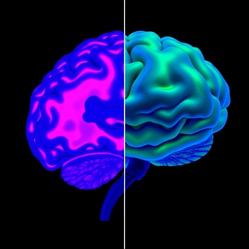
In a groundbreaking escalation of neuroscience research, Mount Sinai’s Living Brain Project has unveiled the most extensive molecular interrogation ever undertaken of the living human brain. This pioneering investigation challenges long-held assumptions about brain biology derived from postmortem samples by demonstrating that living brain tissue exhibits a uniquely distinct molecular signature. By analyzing brain tissue directly from living patients using advanced transcriptomic and proteomic methodologies, the research opens an entirely new paradigm for understanding the brain’s molecular architecture in real-time.
Traditionally, neuroscience and psychiatric disorders have been studied through tissue sourced exclusively from postmortem donations. This has perpetuated the assumption that molecular profiles obtained from deceased brain tissue adequately reflect the molecular state of living brains. However, this assumption remained largely untested due to the rarity and technical complexity of collecting living brain samples. Mount Sinai’s Living Brain Project has now systematically addressed this gap by developing a safe, scalable biopsy technique that harvests small quantities of brain tissue during deep brain stimulation (DBS) surgeries.
Leveraging transcriptomics, which scrutinizes the comprehensive expression of RNA transcripts, alongside proteomics, the large-scale analysis of protein content, the researchers studied approximately 300 samples extracted from the prefrontal cortex of living patients undergoing neurosurgical procedures. The project’s methodological sophistication allowed for intricate comparisons between living brain tissue and standard postmortem samples, exposing profound discrepancies that have important implications for brain science and disease research.
The centerpiece publication in Molecular Psychiatry lays this foundation, providing compelling evidence that gene expression profiles acquired from postmortem brains do not always faithfully mirror the gene expression in living brain tissue. This gap in molecular congruence spans across neurological and psychiatric disease signatures as well as normative phenotypes such as aging. As such, this research cautions against overreliance on postmortem data to model living brain function and pathology without validation.
Alexander W. Charney, MD, PhD, co-leader of the Living Brain Project and Director of The Charles Bronfman Institute for Personalized Medicine, underscores the profound significance of these findings. He calls for an integrative approach to brain research that incorporates the molecular insights gleaned exclusively from living tissue to complement traditional postmortem studies. His remarks emphasize that these revelations enhance, rather than diminish, the value of postmortem research by highlighting the vital, previously inaccessible molecular dimension of living brain tissue.
Expanding upon these insights, the subsequent PLOS ONE publication explores the molecular underpinnings in even greater biochemical depth—specifically, RNA splicing, intron usage, and proteomic variation. This study unveils an extraordinary degree of difference, with over 60% of proteins and a staggering 95% of RNA transcripts processed or expressed differently when comparing living brain tissue with postmortem samples. These disparities emphasize the dynamic and context-dependent nature of brain molecular biology that is lost upon death.
Brian Kopell, MD, Director of Mount Sinai’s Center for Neuromodulation and lead author of the PLOS ONE study, articulates the sheer scale of these differentiated molecular phenomena. He notes that nearly all RNA transcripts examined exhibited altered primary or mature RNA levels or splicing rates in the living brain versus postmortem brain. Importantly, even key interactions between RNA and protein co-expression networks were disrupted in postmortem samples, suggesting that molecular dysregulation post-death goes beyond mere static degradation.
Building on these discoveries, this research advocates for a transformative shift in brain biobanking. With millions worldwide undergoing neurosurgical interventions annually, the feasibility of systematically collecting living brain tissue for diverse biomedical research objectives is within reach. This could catalyze revolutionary advances in deciphering real-time molecular changes related to mood regulation, cognitive processing, and therapeutic responsiveness in a wide array of neuropsychiatric disorders and normal brain functions.
The safety profile of the tissue collection technique developed by the team ensures that brain biopsies can be conducted without compromising patient outcomes, offering a scalable method to construct living brain tissue libraries. Such biobanks would support an unprecedented level of molecular neuroscience inquiry with longitudinal and personalized data sets, reshaping biomedical research trajectories over the decades ahead.
Mount Sinai Health System, the institutional powerhouse behind the Living Brain Project, exemplifies cutting-edge clinical and scientific integration. Housing expansive research infrastructure—including hundreds of clinical and research labs and a vast clinician-scientist workforce—it remains at the vanguard of medical innovation. Their multidisciplinary approach harnesses advances in AI, informatics, and personalized medicine to address complex neurological conditions through a molecular lens.
Beyond the hospital and lab, Mount Sinai’s commitment extends to education and community outreach, ensuring that these transformative discoveries translate into tangible therapeutic breakthroughs and enhanced care paradigms accessible to all patients. The system’s standing—repeatedly validated through top rankings in national and global hospital assessments—affirms its capacity to spearhead bold initiatives like the Living Brain Project.
As neuroscience embraces these revelations, the research community faces a paradigm recalibration. Understanding the living human brain at the molecular level will no longer be extrapolated solely from postmortem proxies. Instead, it will derive directly from the tissue of living subjects, powering forward novel insights into brain health, disease mechanisms, and precision interventions that can drastically improve lives worldwide.
Subject of Research: People
Article Title: A study of gene expression in the living human brain
News Publication Date: 23-Aug-2025
Web References:
https://icahn.mssm.edu/research/friedman/living-brain
https://www.nature.com/articles/s41380-025-03163-1
https://journals.plos.org/plosone/article?id=10.1371/journal.pone.0332651
References:
Molecular Psychiatry (DOI: 10.1038/s41380-025-03163-1)
PLOS ONE (2025 study on RNA splicing and protein expression differences)
Image Credits: Mount Sinai Health System
Keywords:
Molecular neuroscience, Human brain, Proteomics, Omics, Brain, Brain tissue
Tags: deep brain stimulation surgeriesinnovative neuroscience techniquesliving brain biopsiesliving vs. deceased brain samplesmolecular architecture of the brainmolecular differences living brain tissueMount Sinai Living Brain Projectneuroscience research advancementspostmortem brain tissue analysispsychiatric disorders researchreal-time brain tissue studiestranscriptomics and proteomics methodologies


