In a groundbreaking advancement poised to transform the field of gastrointestinal research, a team led by Sobolewski, Planchette, Wójcicki, and colleagues has developed a miniature endoscope capable of capturing high-resolution electrophysiological recordings from the colon of live mice. Published in Nature Communications in 2026, this innovative device promises to unfold new dimensions in the study of neuromuscular regulation and gut physiology, bridging critical gaps in our understanding of gastrointestinal diseases and potential treatments.
The colon, a pivotal organ in the digestive system, is intricately controlled by complex neural circuits and electrophysiological interactions. Until now, direct, high-precision measurement of these electrical signals in vivo has been notoriously difficult due to the organ’s delicate structure, continuous peristaltic movements, and the invasive nature of traditional electrophysiological tools. This miniature endoscope surpasses these hurdles, delivering unprecedented spatiotemporal resolution within the natural physiological environment, paving the way for real-time study of colonic function under both healthy and pathophysiological conditions.
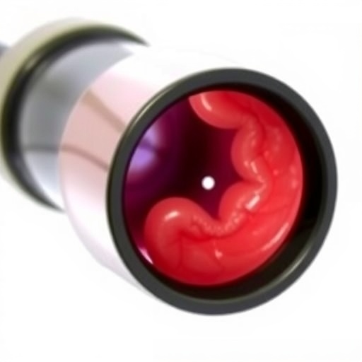
Constructed with cutting-edge microscale fabrication techniques, the miniature endoscope integrates ultra-sensitive microelectrodes within a highly flexible optical fiber. This design ensures minimal invasiveness while maintaining robustness in signal acquisition. The device’s diminutive size not only facilitates access to the narrow lumen of the mouse colon but also ensures compatibility with freely moving subjects, thereby preserving natural gut motility and reducing experimental artifacts that often confound data interpretation in anesthetized or restrained models.
A pivotal aspect of this technology is its dual functionality—merging optical imaging capabilities with electrophysiological sensing in a single platform. The endoscope can simultaneously visualize mucosal morphology and record electrical activity from smooth muscle layers and enteric neurons. This multiplexed capability is a significant technical leap, allowing researchers to correlate real-time structural changes, such as epithelial cell dynamics or inflammation, directly with the electrophysiological signatures that underlie motility patterns and neuronal signaling.
The investigative team undertook a meticulous validation process to establish the fidelity and durability of the device. By implanting the miniature endoscope into live murine models, they monitored spontaneous colonic contractions and evoked neural responses triggered by pharmacological agents. The recorded electrophysiological data exhibited high signal-to-noise ratios and reproducibility, confirming that the device can robustly capture complex electrical patterns inherent to gastrointestinal neural networks without compromising tissue integrity or animal welfare.
From a physiological standpoint, this technology offers a transformative avenue to dissect the enteric nervous system’s functional dynamics. The colon’s neural circuits, often referred to as the “second brain,” play essential roles in regulating digestion, secretion, and motility. Dysfunction in these circuits is implicated in a spectrum of disorders, including irritable bowel syndrome, inflammatory bowel disease, and colorectal cancer. Being able to monitor electrical signaling in vivo opens unprecedented opportunities to decode how neural dysregulation contributes to disease progression, potentially unlocking novel biomarkers and therapeutic targets.
Moreover, the miniature endoscope facilitates longitudinal studies that were previously impractical. Researchers can perform repeated measurements within the same animal over extended periods, allowing for the observation of disease development or the impact of treatment interventions in real time. This capability is particularly valuable for preclinical drug testing, offering detailed insight into how candidate compounds influence neural and muscular physiology within the gut milieu.
The integration of optical and electrophysiological modalities in a single implant also enhances the capacity to study neuroimmune interactions within the colon. Immune cells residing near enteric neurons and epithelial cells can release signaling molecules that modulate electrical activity and motility. The new endoscope’s imaging system can be used to visualize immune cell infiltration or activation while simultaneously recording its electrophysiological consequences, providing a holistic view of gut homeostasis and inflammatory mechanisms.
Technologically, the team addressed significant challenges related to biocompatibility and motion artifacts. The device utilizes advanced biocompatible coatings that minimize tissue inflammation and fibrosis, ensuring stable, long-term recordings. Mechanical stabilization techniques and real-time computational algorithms have been implemented to correct for motion-induced signal distortions caused by peristalsis, which is vital given the inherently dynamic environment of the gastrointestinal tract.
The potential applications of this technology extend beyond basic science. Clinical translation could revolutionize diagnostic endoscopy by introducing electrophysiological assessment alongside visual inspection. This dual assessment could improve early detection of motility disorders or neoplastic changes by identifying abnormal neural patterns that precede gross anatomical alterations. Furthermore, intraoperative use during colorectal surgeries might enhance surgical precision by mapping functional neural zones, reducing the risk of postoperative complications related to nerve damage.
This study also exemplifies the growing trend of miniaturization and multifunctionality in biomedical devices. The researchers’ approach combines advances in microfabrication, flexible electronics, and neurogastroenterology, illustrating the power of interdisciplinary collaboration in tackling long-standing challenges. The robust design principles underpinning this miniature endoscope could inspire similar innovations for studying complex organs such as the heart, bladder, or respiratory tract with matched resolution and physiological relevance.
As researchers continue to refine this technology, future iterations may incorporate wireless data transmission and integrated drug delivery capabilities, enabling seamless interfacing with remote monitoring systems and on-demand therapeutic interventions. The vision of a fully implantable, multifunctional gut probe thus moves closer to reality, promising to unlock unprecedented precision in gastrointestinal diagnostics and therapeutics.
Notably, the insights gleaned from this miniature endoscope align with current priorities in neuroscience and gastroenterology to decode organ-specific neural circuits and their role in systemic homeostasis. Understanding the gut-brain axis has emerged as a critical frontier, given its implications for mental health, metabolic syndromes, and immune regulation. Tools that allow detailed electrophysiological interrogation of the gut in intact, awake subjects fill an essential gap, complementing advances in imaging and molecular biology.
The publication outlines comprehensive experimental protocols and discusses potential future directions, inviting the global scientific community to adopt and advance this platform. By making the device design and software openly accessible, the authors underscore their commitment to collaborative progress, anticipating broad applications not only in research laboratories but also in pharmaceutical development and clinical gastroenterology.
In summary, the creation of this miniature endoscope marks a major milestone in gastrointestinal electrophysiology, offering a unique window into the colon’s living, dynamic neural environment. Its capacity to provide high-resolution, real-time recordings in vivo opens vast possibilities for exploring fundamental physiology, disease mechanisms, and therapeutic strategies. As this technology matures and integrates with complementary innovations, it holds immense promise to reshape our understanding and treatment of digestive health disorders in the years to come.
Subject of Research: Development and application of a miniature endoscope for high-resolution electrophysiological recordings from the colon of live mice.
Article Title: Miniature endoscope for high resolution electrophysiological recordings from the colon of live mice.
Article References:
Sobolewski, A., Planchette, A., Wójcicki, K. et al. Miniature endoscope for high resolution electrophysiological recordings from the colon of live mice. Nat Commun (2026). https://doi.org/10.1038/s41467-026-69144-2
Image Credits: AI Generated
Tags: advancements in gastrointestinal disease treatmentselectrophysiological measurement techniquesgastrointestinal research advancementsgut physiology and diseasehigh-resolution colon recordingsinnovative medical devices for researchmicroscale fabrication in endoscopyminiature endoscope technologyminimally invasive electrophysiologyneuromuscular regulation in the gutoptical fiber integration in medical toolsreal-time study of colonic function

