Cambridge research that has for the first time successfully grown “mini-livers” from adult mouse stem cells has won the UK’s international prize for the scientific and technological advance with the most potential to replace, reduce or refine the use of animals in science (the 3Rs).
Dr Meritxell Huch from the Gurdon Institute, who tonight receives the National Centre for the Replacement, Refinement and Reduction of Animals in Research (NC3Rs) 3Rs Prize, has developed a method that enables adult mouse stem cells to grow and expand into fully functioning three-dimensional liver tissue.
Using this method, cells from one mouse could be used to test 1000 drug compounds to treat liver disease, and reduce animal use by up to 50,000.
Growing hepatocytes (liver cells) in the laboratory has been attempted by liver biologists for many years, since it would reduce their reliance on using mice to study liver disease and would open up new opportunities in medical research and drug safety testing. Until now no laboratory has been successful in deciphering how to isolate and grow these cells.
Liver stem cells are typically found in a dormant state in the liver, only becoming active following injury to produce new liver cells and bile ducts. Dr Huch and colleagues at the Netherlands’ Hubrecht Institute located the specific type of stem cells responsible for this regeneration, which are recognised by a key surface protein (Lgr5+) that they share with similar stem cells in the intestine, stomach and hair follicles.
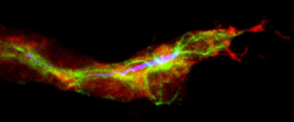
By isolating these cells and placing them in a culture medium with the right conditions, the researchers were able to grow small liver organoids, which survive and expand for over a year in a laboratory environment. When implanted back into mice with liver disease they continued to grow, ameliorating the disease and extending the survival of the mice.
Having further refined the process using cells from rats and dogs, Dr Huch is now moving onto testing it with human cells, which could potentially translate to the development of a patient’s own liver tissue for transplantation.
Commenting on the new method’s potential to reduce animal use in liver research, Dr Huch said: “Typically a study to investigate one potential drug compound to treat one form of liver disease would require up to 50 live animals per experiment, so testing 1000 compounds would need 50,000 mice. By using the liver culture system I developed, we can test 1000 compounds using cells that come from only one mouse, resulting in a significant reduction in animal use.
“If other laboratories adopt this method then the impact on animal use in the liver research field would be immediate. A vast library of potential drug compounds could be narrowed down to just one or two very quickly and cheaply, which can then be tested further in an animal study.”
Dr Vicky Robinson, Chief Executive of the NC3Rs said: “Growing functioning liver cells in culture has been the Holy Grail for liver biologists for many years, so a limitless supply of hepatocytes could have a huge 3Rs impact both on basic research to understand liver disease and for the screening and safety testing of pharmaceuticals. Researchers need to utilise this alternative technology as soon as possible to ensure the benefits to animals and human health are fully realised.”
Professor Kevin Shakesheff, Director of the UK Regenerative Medicine Hub in Acellular Materials, said: “The work of Dr Huch and team demonstrates how three-dimensional culture and molecular biology combine to open new possibilities in the regeneration of complex tissues. The liver is an excellent target for this work as the human body has an ability to regenerate liver tissue that is very hard to replicate in the lab. Unlocking new mechanisms to generate functional liver creates therapeutic approaches for patients with liver disease or injury and could offer a route to high quality human liver models that enhance drug development.”
Story Source:
The above story is based on materials provided by University of Cambridge.


