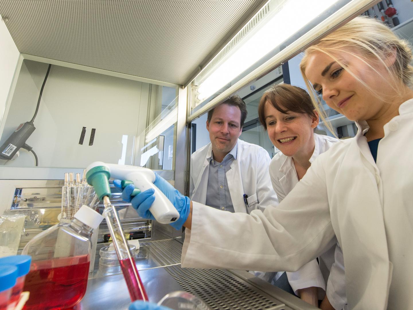
Credit: (c) Photo: Barbara Frommann/Uni Bonn
A new method could push research into developmental brain disorders an important step forward. This is shown by a recent study at the University of Bonn in which the researchers investigated the development of a rare congenital brain defect. To do so, they converted skin cells from patients into so called induced pluripotent stem cells. From these 'jack-of-all-trades' cells, they generated brain organoids — small three-dimensional tissues which resemble the structure and organization of the developing human brain. The work has now been published in the journal Cell Reports.
Investigations into human brain development using human cells in the culture dish have so far been very limited: the cells in the dish grow flat, so they do not display any three-dimensional structure. Model organisms are available as an alternative, such as mice. The human brain has, however, a much more complex structure. Developmental disorders of the human brain can thus only be resembled to a limited degree in the animal model.
Scientists at the Institute of Reconstructive Neurobiology at the University of Bonn applied a recent development in stem cell research to tackle this limitation: they grew three-dimensional organoids in the cell culture dish, the structure of which is incredibly similar to that of the human brain. These "mini brains" offer insight into the processes with which individual nerve cells organize themselves into our highly complex tissues. "The method thus opens up completely new opportunities for investigating disorders in the architecture of the developing human brain," explains Dr. Julia Ladewig, who leads a working group on brain development.
Rare brain deformity investigated
In their work, the scientists investigated the Miller-Dieker syndrome. This hereditary disorder is attributed to a chromosome defect. As a consequence, patients present malformations of important parts of their brain. "In patients, the surface of the brain is hardly grooved but instead more or less smooth," explains Vira Iefremova, PhD student and lead author of the study. What causes these changes has so far only been known in part.
The researchers produced induced pluripotent stem cells from skin cells from Miller-Dieker patients, from which they then grew brain organoids. In organoids, the brain cells organize themselves – very similar to the process in the brain of an embryo: the stem cells divide; a proportion of the daughter cells develops into nerve cells; these move to wherever they are needed. These processes resemble a complicated orchestral piece in which the genetic material waves the baton.
In Miller-Dieker patients, this process is fundamentally disrupted. "We were able to show that the stem cells divide differently in these patients," explains associate professor Dr. Philipp Koch, who led the study together with Dr. Julia Ladewig. "In healthy people, the stem cells initially extensively multiply and form organized, densely packed layers. Only a small proportion of them becomes differentiated and develops into nerve cells."
Certain proteins are responsible for the dense and even packing of the stem cells. The formation of these molecules is disrupted in Miller-Dieker patients. The stem cells are thus not so tightly packed and, at the same time, do not have such a regular arrangement. This poor organization leads, among other things, to the stem cells becoming differentiated at an earlier stage. "The change in the three-dimensional tissue structure thus causes altered division behavior," says Ladewig. "This connection cannot be identified in animals or in two-dimensional cell culture models."
The scientist emphasizes that no new treatment options are in sight as a result of this. "We are undertaking fundamental research here. Nevertheless, our results show that organoids have what it takes to herald a new era in brain research. And if we better understand the development of our brain, new treatment options for disorders of the brain can presumably arise from this over the long term."
###
Publication: Vira Iefremova, George Manikakis, Olivia Krefft, Ammar Jabali, Kevin Weynans, Ruven Wilkens, Fabio Marsoner, Björn Brändl, Franz-Josef Müller, Philipp Koch and Julia Ladewig: An Organoid-Based Model of Cortical Development Identifies Non-Cell-Autonomous Defects in Wnt Signaling Contributing to Miller-Dieker Syndrome; Cell Reports; DOI: 10.1016/j.celrep.2017.03.047
Contact:
Dr. Julia Ladewig,
Institute of Reconstructive Neurobiology
University of Bonn
Tel. +49-228-6885547
E-mail: [email protected]
Media Contact
Dr. Julia Ladewig
[email protected]
49-228-688-5547
@unibonn
http://www.uni-bonn.de


