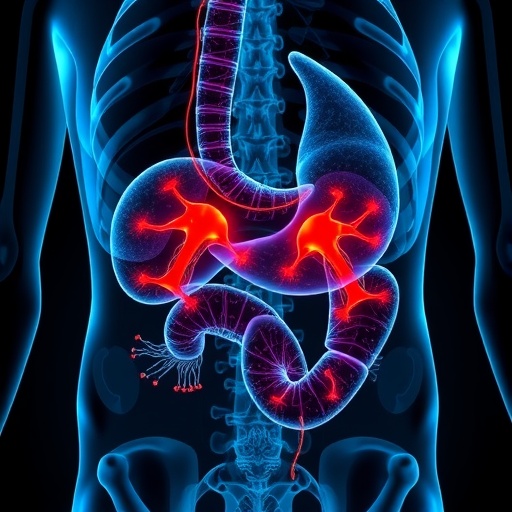
In a groundbreaking study set to redefine our understanding of cancer metastasis, researchers have unveiled the intricate spatiotemporal dynamics of necroptosis within the progression of gastric cancer, focusing particularly on the mechanisms that drive lymph node metastasis. The investigation, employing cutting-edge single-cell and spatial transcriptomic technologies, provides an unprecedented cellular-level dissection of the evolving tumor microenvironment and the role that programmed necrotic cell death plays in facilitating cancer dissemination.
Gastric cancer remains one of the most lethal malignancies worldwide, primarily due to its aggressive nature and the propensity for early metastasis to regional lymph nodes. The molecular and cellular pathways underlying this metastatic spread have remained elusive, complicating efforts to develop targeted therapies. This latest research offers pivotal insights by tracking necroptosis—an inflammatory form of regulated cell death—over time and space within the tumor milieu, revealing how necroptotic signaling cascades may orchestrate the metastatic process.
Using sophisticated single-cell RNA sequencing alongside spatial transcriptomics, the scientists were able to resolve the heterogeneity among tumor and stromal cells with unparalleled resolution. This dual approach allowed them to map the temporal evolution of necroptotic events and identify distinct cellular subpopulations that appear to drive lymph node colonization. These necroptotic niches were characterized not just by dying cells but by an active interplay between immune components, endothelial cells, and cancer stem-like cells, painting a complex picture of microenvironmental remodeling.
One of the most striking revelations from the study is the demonstration that necroptosis is not merely a terminal phenomenon but functions dynamically to promote metastatic competence. Necroptotic cells release specific damage-associated molecular patterns (DAMPs) and cytokines, which were observed to modulate the trafficking and activation status of immune cells in the tumor vicinity. This inflammatory milieu facilitates the breakdown of extracellular matrix barriers and enhances the invasiveness of cancer cells, thereby accelerating their escape into lymphatic vessels.
Moreover, the temporal profiling indicated that necroptosis spikes during critical windows of tumor-host interaction, particularly preceding lymphatic invasion. This suggests a carefully choreographed sequence where necroptotic signaling primes the microenvironment for metastatic dissemination. The spatial data further corroborated these findings, showing hotspots of necroptosis aligned with areas of heightened lymphangiogenesis and immune infiltration, underscoring a spatially restricted, yet systemically impactful, process.
The involvement of necroptosis in such a pivotal step of cancer progression underscores its dualistic nature—traditionally viewed as a tumor-suppressing mechanism due to its cell-killing potential, it paradoxically appears to facilitate tumor spread under certain conditions. This nuanced understanding challenges previous dogmas and opens new therapeutic avenues where modulation of necroptotic pathways could switch this deadly signal into a therapeutic vulnerability.
Further characterization revealed that key necroptosis regulators, such as RIPK1, RIPK3, and MLKL, exhibit altered expression patterns in metastatic lesions compared to primary tumors. These molecules orchestrate the necroptotic cascade and are potential candidates for targeted intervention. The study’s findings propose that inhibiting these orthodox mediators could disrupt the pro-metastatic signaling loops, thereby stalling lymph node colonization and ultimately improving patient outcomes.
The role of the immune system, a recurrent theme in modern oncological research, is intricately woven into the necroptotic narrative portrayed here. Immune subpopulations, including tumor-associated macrophages and cytotoxic T cells, were found in close proximity to necroptotic foci, suggesting a complex cross-talk that may either facilitate immune evasion or provoke anti-tumor immunity depending on context and timing. This revelation holds promise for designing immunomodulatory therapies tailored to the necroptotic landscape of a patient’s tumor.
Importantly, this research leverages the strength of spatial transcriptomics to transcend the limitations of bulk analyses, which often obscure cellular heterogeneity and spatial context. By anchoring gene expression data to actual tissue architecture, the study elucidates how microenvironmental cues are spatially coordinated with cellular fate decisions—particularly necroptosis—and how this orchestration drives metastatic success.
Adding to its impact, the study underscores the utility of integrating single-cell and spatial biology as a gold standard in unraveling cancer complexity. This integrative methodology paves the way for future studies to explore similar mechanisms in other cancer types, potentially uncovering universal or cancer-specific necroptotic signatures associated with metastasis.
While the translational applications of these findings are still emerging, the identification of necroptosis as a critical driver of lymph node metastasis invites the design of novel diagnostic tools. Biomarkers derived from necroptotic signaling components could serve as prognostic indicators or as predictors of response to emerging targeted therapies aiming to disrupt necroptosis-induced metastasis.
This research does not only enrich basic cancer biology but also resonates with the clinical challenge of managing lymph node metastasis—a primary determinant of patient prognosis and therapeutic strategy in gastric cancer. By shining a light on the temporal and spatial evolution of necroptosis, the work informs surgical decisions, adjuvant therapy regimens, and surveillance protocols, potentially transforming clinical workflows.
The study’s multidisciplinary approach combines molecular biology, genomics, immunology, and spatial analysis to construct a comprehensive atlas of necroptosis-mediated metastatic evolution. This atlas serves both as a resource and a roadmap for researchers aiming to dissect the layered complexity of tumor progression from a cellular and spatial vantage point.
In conclusion, this pioneering investigation by Hu, Shen, Zhang, and colleagues marks a paradigm shift in our understanding of tumor biology. By elucidating how necroptosis, a cell death modality once considered merely destructive, actively propels lymph node metastasis in gastric cancer, it charts a new frontier for cancer research and therapeutic innovation. As the community builds on these insights, the ultimate beneficiaries will be the patients, who may one day receive treatments precisely calibrated to intercept necroptotic signaling and prevent cancer’s deadly spread.
Subject of Research: Necroptosis mechanisms driving lymph node metastasis in gastric cancer
Article Title: Single-cell and spatial dissection of necroptosis spatiotemporal evolution driving lymph node metastasis in gastric cancer
Article References:
Hu, Y., Shen, F., Zhang, H. et al. Single-cell and spatial dissection of necroptosis spatiotemporal evolution driving lymph node metastasis in gastric cancer. Cell Death Discov. 11, 535 (2025). https://doi.org/10.1038/s41420-025-02815-z
Image Credits: AI Generated
DOI: 17 November 2025
Tags: cancer metastasis mechanismscellular heterogeneity in tumorsinflammatory cell death in cancerlymph node metastasis in gastric cancermetastatic spread of cancernecroptosis in gastric cancernecroptotic signaling pathwaysprogrammed necrotic cell deathsingle-cell RNA sequencing applicationsspatial transcriptomics technologytargeted therapies for gastric cancertumor microenvironment dynamics


