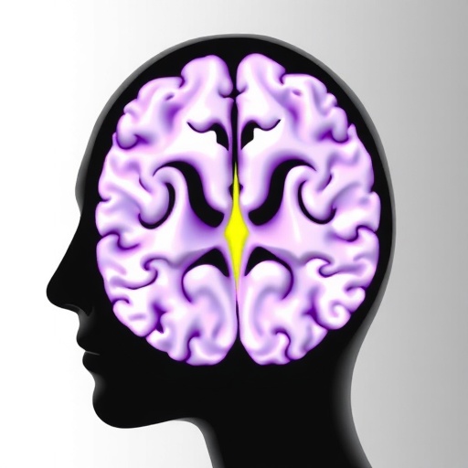
In a groundbreaking advancement for neuroscience, researchers have unveiled an unprecedented cortical and subcortical map of the human allostatic–interoceptive system utilizing ultra-high-field 7 Tesla functional magnetic resonance imaging (fMRI). This innovative study captures the intricate and dynamic interplay between brain regions responsible for internal bodily regulation and external environmental adaptation, highlighting a complex network that governs how humans maintain homeostasis amid changing conditions. By achieving exquisitely detailed imaging at the submillimeter scale, the team illuminates the neural architecture underpinning the brain’s capacity to sense and regulate the body’s internal state, a feat previously beyond the reach of neuroimaging technologies.
The human allostatic–interoceptive system, central to sustaining physiological equilibrium, integrates signals from internal organs and the external environment to guide brain-mediated adjustments in behavior, metabolism, and cardiovascular function. The term allostasis refers to the brain’s process of achieving stability through physiological or behavioral change, an extension of the classical concept of homeostasis. Interoception, meanwhile, is the sensory process by which the brain receives and interprets physiological feedback from within the body, encompassing visceral sensations like heartbeat, respiration, and gut activity. Understanding this integrative system is crucial for elucidating how the brain maintains health, adapts to stress, and how its dysfunction contributes to a vast array of neuropsychiatric and systemic diseases.
Leveraging the unparalleled spatial resolution and sensitivity of 7 Tesla MRI scanners, the research team was able to precisely map cortical and subcortical areas implicated in allostatic and interoceptive processing. The 7 Tesla field strength permits detection of subtle blood oxygen level-dependent (BOLD) signals from minute brain structures that house specialized neurons, previously difficult to image due to technical constraints and signal noise. By combining task-based and resting-state fMRI paradigms, the researchers systematically charted intrinsic connectivity and activation patterns associated with interoceptive awareness and autonomic control.
Among the landmark observations is the delineation of distinct cortical hubs within the insular cortex, anterior cingulate cortex, and ventromedial prefrontal cortex, each exhibiting unique but interconnected roles in processing interoceptive signals and orchestrating adaptive physiological responses. The posterior insula emerged as a primary cortical entry point for visceral afferent information, receiving inputs from lamina I of the spinal cord and vagal nerve pathways, thus acting as a visceral sensory cortex. Conversely, anterior insular regions synthesized these physiological inputs with emotional and cognitive contextual information, underpinning interoceptive awareness and subjective feeling states.
Subcortically, the study reveals nuanced participation of deep brain structures such as the hypothalamus, periaqueductal gray (PAG), and brainstem nuclei in modulating autonomic and endocrine functions integral to allostasis. The hypothalamus, a pivotal allostatic coordinator, integrates afferent viscerosensory inputs and orchestrates neuroendocrine output to regulate stress responses, metabolism, and circadian rhythms. Notably, the periaqueductal gray was implicated in modulating pain and defensive behaviors linked to homeostatic threat, while discrete brainstem centers fine-tuned sympathetic and parasympathetic outflow, accounting for moment-to-moment adjustments in heart rate and respiration.
This comprehensive mapping also identified circuitry involving the amygdala and thalamus as critical relays for integrating visceral sensory data with emotional salience and cognitive appraisal. The amygdala’s engagement in interoceptive processing highlights its role in encoding bodily signals that underpin affective states and decision-making. Meanwhile, thalamic nuclei were found to facilitate communication between brainstem inputs and higher cortical areas, establishing a bidirectional pathway essential for conscious perception of internal states.
One of the most revolutionary elements of the research is the fusion of cortical and subcortical maps to illustrate how these distributed regions form an integrated network functioning as an allostatic-interoceptive axis. Through advanced connectivity analyses, the researchers demonstrated bidirectional coupling and hierarchical processing cascades that fine-tune physiological set points in anticipation of environmental demands. This dynamic network supports predictive coding models where the brain continuously generates and updates hypotheses about bodily states, thus minimizing prediction errors by adjusting autonomic outputs.
The study’s insights extend beyond basic neuroscience, holding transformative implications for understanding psychiatric conditions like anxiety, depression, and substance use disorders where dysregulated interoception and allostatic load are prominent features. For example, heightened interoceptive sensitivity or impaired insular integration may contribute to abnormal anxiety responses or mood dysregulation. By pinpointing the precise locations and functional connectivity profiles of these allostatic hubs, targeted neuromodulation therapies such as transcranial magnetic stimulation or deep brain stimulation may be refined to restore healthy brain-body communication.
Moreover, this cutting-edge approach sets a new standard for high-resolution functional mapping in humans, opening pathways for longitudinal studies tracing how these allostatic-interoceptive networks evolve across the lifespan or in response to treatments. It also prompts a re-evaluation of the traditional brain-centric approach to neurobehavioral disorders, foregrounding the importance of somatic markers and body-based predictive systems as central etiological factors.
Technically, the success of this endeavor hinged on overcoming formidable challenges inherent in 7 Tesla fMRI, including susceptibility artifacts caused by magnetic field inhomogeneities, physiological noise from cardiac and respiratory cycles, and motion artifacts. The research team employed sophisticated preprocessing pipelines incorporating distortion correction, motion correction, and physiological noise modeling to preserve signal fidelity. Furthermore, they designed novel interoception-evoking experimental paradigms that engaged cardiovascular, respiratory, and visceral sensory inputs while participants remained in the scanner, enabling concurrent imaging of brain-wide responses to authentic internal stimuli.
With this exquisitely detailed atlas of the allostatic-interoceptive system now publicly available, the neuroscience community gains a foundational resource for integrating brain physiology with peripheral organ systems. This integrative framework will catalyze cross-disciplinary efforts bridging neuroscience, endocrinology, immunology, and psychology, fostering a holistic understanding of how brain and body maintain equilibrium amid constant flux. The work exemplifies how advances in neuroimaging technology combined with conceptual rigor can unravel the complex adaptive machinery governing human health.
Looking forward, expanding this mapping to clinical populations will be paramount. Delineating deviations in allostatic-interoceptive circuitry in disorders such as hypertension, irritable bowel syndrome, chronic pain, and post-traumatic stress disorder could reveal biomarkers predictive of disease progression or treatment response. Additionally, synergizing ultra-high-resolution imaging with emerging modalities like molecular neuroimaging or simultaneous multi-organ physiological monitoring could deepen multimodal representations of mind-body integration.
In summary, this landmark study represents a paradigm shift, charting a comprehensive neurofunctional atlas of the human brain’s allostatic and interoceptive machinery at remarkable granularity. It illuminates the neural substrates by which the brain monitors and regulates the internal milieu, maintaining adaptive homeostasis amidst ever-changing internal and external environments. By transcending previous technical limitations and conceptual boundaries, it sets a vibrant agenda to decipher the enigma of embodied cognition and somatic regulation in health and disease. This achievement underscores the power of high-field neuroimaging and integrative neuroscience to illuminate the brain’s role as a master regulator of the living organism.
Subject of Research:
Mapping the human allostatic–interoceptive system in the brain using ultra-high-field 7 Tesla fMRI to understand neural substrates of internal bodily regulation and adaptive physiological processes.
Article Title:
Cortical and subcortical mapping of the human allostatic–interoceptive system using 7 Tesla fMRI
Article References:
Zhang, J., Chen, D., Deming, P. et al. Cortical and subcortical mapping of the human allostatic–interoceptive system using 7 Tesla fMRI. Nat Neurosci (2025). https://doi.org/10.1038/s41593-025-02087-x
Image Credits:
AI Generated
Tags: allostasis and physiological stabilitybrain regions in homeostasiscortical and subcortical mappingdynamic interplay between brain and bodyhuman allostatic-interoceptive systeminternal and external environmental adaptationinteroception and visceral sensationsneural architecture of bodily regulationneuroimaging advancements in neurosciencephysiological equilibrium in human biologyultra-high-field 7 Tesla fMRIunderstanding brain health and stress response


