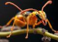
In a groundbreaking advance for biological sciences, a consortium of researchers from the Okinawa Institute of Science and Technology (OIST) and the Karlsruhe Institute of Technology (KIT), along with global collaborators, has unveiled an unprecedented database comprising over 2000 high-resolution 3D models of ants. This revolutionary project, known as Antscan, leverages state-of-the-art synchrotron-powered micro-computed tomography (micro-CT) technology to capture the intricate morphology of ants at micrometer resolution, not only preserving the external exoskeletons but also exposing detailed internal structures such as musculature, neural networks, digestive tracts, and stinger apparatuses.
Understanding the form and structure of organisms has always been fundamental to biology, yet the sheer complexity and minuteness of many species, particularly insects, have long posed formidable challenges for comprehensive morphological studies. Traditional techniques rely heavily on specimen mounting and preservation methods that maintain external features while internal anatomy rapidly degrades post-mortem. Moreover, conventional imaging tools such as laboratory-based CT scanners are prohibitively slow and costly for mass-scaling research, often restricting studies to individual or few specimens.
Antscan transcends these constraints by employing a synchrotron particle accelerator as a powerful X-ray source coupled with automated robotic sample handling to facilitate ultra-high-throughput scanning. By orchestrating a robotic arm to systematically exchange specimen vials, the system generates approximately 3000 two-dimensional X-ray projections per specimen. Advanced computational algorithms then reconstruct these projections into comprehensive 3D tomograms, revealing ant morphology with unmatched resolution and fidelity. Remarkably, this technology condensed what would conventionally require six years of continuous scanning down to a single week.
The breadth and depth of Antscan’s dataset are transformative for entomologists, morphologists, and the broader scientific community. Every specimen’s metadata—including taxonomy, collection locality, collector identity, and caste designation—has been meticulously standardized to ensure data integrity and facilitate comparative research. The collaboration spanned a network of museums, research institutions, and private collections worldwide, highlighting the project’s global scale and ambition.
One of the most striking facets of Antscan is its commitment to open science. All raw data are freely accessible through an online portal featuring a built-in 3D viewer, democratizing access to what has hitherto been an extraordinarily resource-intensive form of data generation. This accessibility extends opportunities beyond academia, inviting citizen scientists, educators, digital artists, and conservationists to explore and utilize the models for diverse purposes. The detailed visualizations of musculature and articulation open new avenues in biomechanical modeling, enhancing our understanding of insect locomotion and physiology.
From an ecological perspective, ants serve as keystone organisms in numerous terrestrial ecosystems, their social and structural diversity underpinning complex environmental interactions. The Antscan project not only provides a reservoir of morphological data but also paves the way for integrative studies combining genomics, biodiversity mapping, and phenotypic variation. Previous complementary efforts by OIST have already produced global distribution maps of ant biodiversity and high-quality genome assemblies covering most ant genera, setting the stage for multifaceted investigations.
The synergy of big data and advanced imaging realized by Antscan exemplifies the dawning era of quantitative phenomics, where organismal shape and form can be studied at scales and resolutions previously unimaginable. Professor Evan Economo, a co-leader of the project, highlights how this initiative advances a paradigm shift, facilitating the capture, analysis, and sharing of morphological data with unprecedented breadth. The potential for cross-disciplinary insights that merge evolutionary biology, ecology, and computational modeling is immense.
Technically, the integration of synchrotron-based micro-CT scanning into biological research represents a novel application of physics and engineering resources long dominated by materials science and medical imaging. The method involves exposing specimens to a highly collimated, intense X-ray beam, capturing slices at micron-scale increments. The resultant volumetric data enable virtual dissection and exploration without physical damage, preserving valuable specimens for future study while maximizing information yield.
Moreover, the operation’s high-throughput nature required innovations in robotics and software automation. Sorting and cataloging over 2000 ant specimens demanded coordinated efforts involving numerous experts who painstakingly classified ants by species and caste before scanning. The realized workflow sets a new benchmark for mass phenotyping projects, suggesting that parallel efforts could soon be undertaken for other taxonomically rich and morphologically complex groups.
The implications of this work extend to ecological and evolutionary research realms. In a related study published in Science Advances, Antscan data were instrumental in investigating the balance between worker ant quantity and quality within colonies. Findings indicate that favoring numerous nutritionally inexpensive workers over fewer, heavily armored individuals correlates with the evolution of larger and more resilient social systems—a revelation made possible by the rich morphological detail Antscan provides.
Looking forward, Antscan’s open-access framework promises to catalyze myriad innovative applications, spanning from biomimetic robotics design, ecological monitoring, species identification, to public engagement in biodiversity sciences. By lowering barriers to high-resolution morphological data, this initiative may inspire new models of collaborative research and education, galvanizing a broad community invested in understanding and preserving the diversity of life forms on our planet.
In sum, Antscan is a landmark achievement in phenomics, marrying cutting-edge technology and collaborative science to unlock the detailed morphology of some of Earth’s most ecologically significant insects. This resource not only enriches fundamental biological knowledge but also exemplifies how modern techniques can overcome historical bottlenecks, enabling comprehensive, scalable, and accessible study of organismal form and function.
Subject of Research: Animals
Article Title: High-throughput phenomics of global ant biodiversity
News Publication Date: 5-Mar-2026
Web References:
Antscan project portal: https://antscan.info/
Related global ant biodiversity map: https://www.oist.jp/news-center/news/2022/8/4/new-global-map-ant-biodiversity-reveals-areas-may-hide-undiscovered-species
Ant genomes publication: https://www.cell.com/cell/fulltext/S0092-8674(25)00617-8
Study on ant colony organization: https://www.oist.jp/news-center/news/2025/12/20/ant-societies-rose-trading-individual-protection-collective-power
References:
Katzke, J. et al. (2026). High-throughput phenomics of global ant biodiversity. Nature Methods, DOI: 10.1038/s41592-026-03005-0.
Image Credits: Thomas van de Kamp
Keywords: Antscan, Ant morphology, Micro-CT scanning, Synchrotron imaging, Phenomics, Biodiversity, 3D modeling, Insect anatomy, High-throughput imaging, Big data in biology, Computational morphology, Ant ecology
Tags: 3D ant morphology databaseadvanced biological imaging techniquesant anatomical structuresautomated robotic sample handlingglobal ant diversity mappinghigh-resolution insect imaginginnovative taxonomic research toolsinternal anatomy of antsmass-scale insect morphology studymicrometer-resolution biological imagingneural network visualization in antssynchrotron micro-CT scanning

