In a groundbreaking study poised to redefine our understanding of Alzheimer’s disease (AD) pathology, researchers have leveraged cutting-edge magnetic resonance microscopy (MRM) to chart, with exceptional precision, the widespread structural alterations occurring throughout the brain in a well-established mouse model of AD. The investigation, conducted by Tian, Hornburg, Austin, and colleagues, systematically scrutinizes the morphological changes across 231 distinct brain regions in 14-month-old 5×FAD BXD hybrid mice, illuminating previously uncharted realms of neural vulnerability and resilience.
Alzheimer’s disease, characterized primarily by progressive cognitive decline and neurodegeneration, is notorious for its insidious impact on neural circuitry and brain morphology. However, past studies have often been constrained to examining limited brain regions or gross morphological changes. This new research overcomes such limitations by employing diffusion tensor magnetic resonance imaging (DT-MRI) at a remarkable 25-micron resolution, thereby enabling a comprehensive and highly granular mapping of the disease’s impact across the forebrain and brainstem. This unprecedented spatial resolution permits an intricate dissection of the volumetric and microstructural brain alterations resulting from mutations in the amyloid precursor protein (APP) and presenilin 1 (PSEN1) genes—two key players in AD pathogenesis.
Upon analyzing the data, the researchers observed that more than half of the 231 brain regions exhibited significant volume changes. Intriguingly, these alterations were not uniformly degenerative; some brain areas demonstrated unexpected volumetric expansion rather than shrinkage. The neocortex, hippocampus, and amygdala—regions crucial for cognition, learning, and emotional regulation—manifested swelling of up to 10%, defying conventional expectations of atrophy. This focal hypertrophy challenges prevailing assumptions about the straightforward nature of AD-related neurodegeneration and suggests that the disease’s impact is more multifaceted and nuanced.
Contrasting the expansion in certain regions, the study identified considerable shrinkage within the thalamus, brainstem, and the majority of white matter tracts. These findings underscore a spatially heterogeneous pattern of neurodegeneration, where neural systems involved in sensory integration and basic autonomic functions could be disproportionately compromised. Despite these extensive local changes, the total brain volume in mutant mice remained remarkably stable, indicating that volume loss in some areas might be offset by gains in others, or potentially by neuroinflammatory or gliotic processes leading to swelling.
One of the most compelling facets of this research is the pronounced sex-specific differences in structural variability. Female mice exhibited greater variance in the volume of individual brain regions compared to males, implying that the neuroanatomical consequences of AD mutations may be modulated by sex-dependent biological factors. This finding resonates with clinical observations in humans, where the prevalence and progression of Alzheimer’s disease differ between men and women, yet the underlying neurobiological substrates have remained elusive.
Beyond structural mapping, the team integrated behavioral analyses focusing on fear acquisition and contextual memory performance, vital cognitive domains affected by AD. The volumetric changes in several brain regions were found to covary robustly with these behaviors, and perhaps most intriguingly, the directionality of these correlations diverged between mutant and control mice. This means that a brain region enlarging in an AD model might relate to worsened memory there, whereas the same region’s size in a healthy brain might predict better memory function, pointing to a complex and context-dependent relationship between neuroanatomy and behavior in the disease state.
Technically, the use of diffusion tensor MRI allowed characterization not only of volumetric changes but also inferred alterations in tissue microarchitecture, providing insights into the integrity of neural pathways and fiber tracts. White matter degeneration in Alzheimer’s models is a well-documented phenomenon, often associated with cognitive decline, and this study’s detailed tract-specific shrinkage highlights potential targets for therapeutic intervention aiming to preserve or restore white matter health.
The choice of the 5×FAD BXD hybrid strain is noteworthy because it carries five familial AD mutations and exhibits significant amyloid pathology by 14 months of age, closely mimicking human disease progression. Employing this genetically diverse background enhances the translational relevance of the findings by capturing variability more akin to that encountered in human populations, including genotype-by-environment interactions.
The implications of mapping 231 discrete regions across rostrocaudal (front-to-back) and mediolateral (side-to-side) brain axes extend well beyond morphological description. It allows a multidimensional atlas of Alzheimer’s-associated neurodegeneration that can facilitate precision targeting of brain areas for future interventions. Moreover, the counterintuitive regional swelling observed offers fertile ground for exploring whether such hypertrophies represent pathological hallmarks, neuroinflammatory responses, or compensatory neuroplasticity.
This comprehensive morphological benchmark not only enriches our mechanistic understanding of AD but also establishes a powerful framework for evaluating the efficacy of emerging therapeutics in preclinical trials. By providing a detailed and quantitative baseline of structural changes linked to genotype and behavior, future studies can discern how candidate drugs or interventions mediate brain-wide effects, potentially identifying early biomarkers of treatment response before overt clinical improvements manifest.
Furthermore, these findings open avenues to investigate how molecular cascades underlying APP and PSEN1 mutations differentially influence discrete neuroanatomical structures and circuits, integrating multiomics approaches with imaging phenotypes. Such cross-modal investigations hold promise for unraveling the cascading events from genetic mutation to cellular dysfunction, circuit disruption, and ultimately cognitive deficits.
This work exemplifies the power of high-resolution neuroimaging in animal models to decipher the complex neurobiology of Alzheimer’s disease. It underscores that AD is not simply a disease of uniform atrophy but a disorder involving intricate spatial patterns of degeneration and growth, interacting dynamically with behavior and sex. These insights reinforce the notion that effective AD therapeutics will need to be multifactorial, aimed at preserving or restoring the delicate balance of structural and functional brain integrity.
The established atlas serves as a valuable resource for the neuroscience community, offering a comprehensive dataset for benchmarking and comparative studies with other neurodegenerative models or aging paradigms. It also provides a crucial reference for novel imaging biomarker development, paving the way for more sensitive diagnostic and prognostic tools in clinical settings.
As the field advances, the integration of these detailed structural maps with longitudinal cognitive assessments and molecular analyses will be essential to track disease trajectories and identify windows of intervention. The current study lays the essential groundwork for such longitudinal investigations, underscoring the value of systematic, whole-brain analyses over piecemeal approaches.
In summary, this landmark study combines state-of-the-art imaging technology with sophisticated genetic and behavioral paradigms to chart the landscape of Alzheimer’s disease in an unprecedentedly comprehensive manner. By revealing region-specific hypertrophy and atrophy, sex-dependent variability, and complex neurobehavioral relationships, it sets a new standard for preclinical AD research and opens transformative pathways toward understanding and treating this devastating disorder.
Subject of Research: Magnetic resonance microscopy mapping of brain structural changes in a genetic mouse model of Alzheimer’s disease.
Article Title: Magnetic resonance microscopy maps widespread effects of Alzheimer’s disease on brain structures and behavior in mice.
Article References:
Tian, Y., Hornburg, K., Austin, W. et al. Magnetic resonance microscopy maps widespread effects of Alzheimer’s disease on brain structures and behavior in mice. Nat Neurosci (2026). https://doi.org/10.1038/s41593-025-02199-4
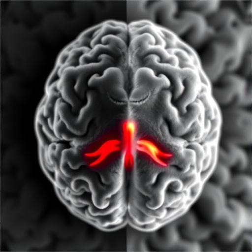
Image Credits: AI Generated
DOI: https://doi.org/10.1038/s41593-025-02199-4
Tags: 5×FAD BXD mouse model Alzheimer’s studyadvanced imaging techniques for cognitive declineamyloid precursor protein mutation effectsbrain volumetric changes in Alzheimer’s diseasecomprehensive brain region analysis in ADdiffusion tensor MRI for brain morphologyhigh-resolution brain imaging in neurodegenerationmagnetic resonance microscopy in Alzheimer’s researchmapping neural vulnerability in Alzheimer’smicrostructural brain alterations in neurodegenerative disorderspresenilin 1 gene role in Alzheimer’sstructural brain changes in mouse models


