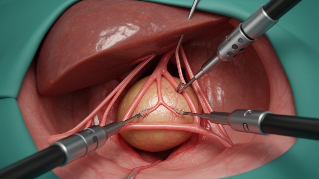
Laparoscopic cholecystectomy remains a cornerstone of modern general surgery, yet the procedure continues to be associated with a persistent risk of bile duct injury, an event that can have profound consequences for patient outcomes and quality of life. This risk is frequently compounded by anatomical variability, inflammation, or dense adhesions, which often render conventional surgical landmarks like Calot’s triangle or the traditional Critical View of Safety technically challenging to establish accurately. To address these persistent hurdles, surgeons have developed a novel dissection strategy known as the Madany’s Triangle technique. This approach serves as a dynamic, safety-oriented framework designed to enhance the visualization of critical biliary structures during surgery, potentially providing a more adaptable method for surgeons operating in complex or inflamed environments.
The traditional approach to biliary surgery relies heavily on fixed anatomical landmarks that are often present before dissection commences, which can be misleading in the presence of severe cholecystitis or anatomical variations. In contrast, the Madany’s Triangle is defined as a dynamic surgical space that only emerges after a series of precise, staged dissection steps have been completed. This technique is predicated on the early skeletonization of the proximal third of the gallbladder, the infundibulum, and the cystic duct, alongside the early scarification of the cystic artery. By prioritizing these specific movements, the surgeon creates a clearer window that is shifted away from the common bile duct and the right hepatic artery, thereby minimizing the risk of inadvertent damage to these vital structures during the routine steps of the operation.
To achieve the Madany’s Triangle, the surgeon utilizes the dynamic nature of the surgical field, which changes significantly depending on the direction of traction applied to the Hartmann’s pouch and the specific angle of the laparoscopic camera. From a left-side view, applying traction to the Hartmann’s pouch toward the right creates tension that reveals the triangle. The lateral border is defined by the skeletonized cystic duct, while the superior border is formed by the posterior surface of the skeletonized proximal third of the gallbladder. The medial border is delineated by an imaginary line connecting the junction of the cystic duct with the common bile duct to the anterior end of the dissected cystic plate. This structured visualization provides a clear checkpoint that must be met before any definitive clipping or cutting occurs.
A complementary perspective is obtained from the right-side view, which becomes apparent when traction is redirected to the left and the scope is repositioned. In this configuration, the medial border is defined by the dissected cystic duct, the superior border remains the posterior surface of the skeletonized proximal third of the gallbladder, and the lateral border is the line from the cystic duct–common bile duct junction to the anterior end of the dissected cystic plate. The ability to manipulate the visibility of this triangular window through controlled traction demonstrates the method’s reliance on real-time anatomic confirmation rather than static, predefined assumptions. This feedback mechanism serves as a crucial safety tool, ensuring that if the triangle is not clearly visible, the surgeon has not yet achieved the necessary level of dissection to proceed safely.
The procedural methodology follows a strict, step-by-step sequence designed to maximize exposure. The process begins with the dissection of the peritoneal attachment on both the right and left sides of the proximal gallbladder, proceeding from proximal to distal on the right and distal to proximal on the left. Following this, the surgeon releases the cystic artery, an act described as freeing the artery from its “seatbelt effect.” Because the cystic artery is typically shorter than the cystic duct, its release allows the often tortuous or S-shaped duct to straighten, significantly widening the working field and making the anatomy easier to interpret. Subsequent steps involve thorough skeletonization of the posterior surface of the gallbladder until the plane from the right and left meet, effectively creating a clean surgical window.
Once the posterior dissection is complete, the surgeon focuses on the careful isolation of the cystic duct. Dissection of the duct from both the left and right sides continues until the junction with the common bile duct is clearly identified. This attention to detail at the distal cystic duct is an essential component of the Madany’s approach, specifically aimed at preventing thermal injury and avoiding the creation of an excessively long cystic duct stump. By ensuring these structures are skeletonized and exposed before any clipping, the surgeon forces themselves to achieve the Critical View of Safety criteria consistently. The technique does not replace existing safety protocols but rather operationalizes them through a more rigorous and repeatable dissection sequence.
Clinical observations derived from a series of 238 patients who underwent this technique between December 2019 and March 2023 provide preliminary evidence of its feasibility. Within this cohort, the female-to-male ratio was 5:1, and the most common indication for surgery was chronic cholecystitis, representing approximately 66.8% of the cases. The average operative time was reported at 63 minutes. While this experience-based technical note lacks the formal statistical analysis and control groups found in prospective, randomized clinical trials, surgeons applying the technique have reported significant subjective improvements. These include clearer anatomical definition, a higher degree of confidence during the dissection of challenging porta hepatis areas, and a perceived reduction in the need for conversion to open surgery.
One of the primary advantages cited by the developers is the technique’s ability to minimize the risks associated with misidentification of the common bile duct, which remains one of the leading causes of biliary injury. By shifting the dissection zone laterally and anteriorly, the technique provides a safer distance from the major bile ducts. Furthermore, unlike the fundus-first technique, which can limit the ability to retract the liver effectively, the Madany’s Triangle method maintains the integrity of the gallbladder fundus for retraction, which is vital for exposure. This combination of early, wide exposure and controlled, skeletonized dissection allows for a clearer view of biliary anomalies without necessarily relying on deep, high-risk maneuvers near the porta hepatis.
Despite these clinical benefits, the technique carries inherent limitations that warrant cautious interpretation. Like the traditional Critical View of Safety, success is highly dependent on the skill and experience of the operating surgeon, particularly when navigating near the porta hepatis. The early phase of adoption can also be associated with increased operative time as surgeons focus on the meticulous dissection required to build the triangle dynamic. Additionally, in cases of severe acute cholecystitis characterized by dense adhesions or distorted anatomical structures, such as a missing or extremely short cystic duct, achieving the necessary exposure to form the triangle may not be feasible, requiring surgeons to revert to established bailout strategies.
The current lack of large-scale, multicenter, randomized controlled trials means that while the Madany’s Triangle shows promise, its clinical superiority over conventional methods remains to be validated. The dynamic nature of the triangle also requires precise coordination between the camera position and trocar angulation, meaning that operator experience directly impacts the reproducibility of the visual landmarks. Consequently, the technique should currently be viewed as an evolutionary framework that complements, rather than supplants, established standards. Future research aimed at comparing this method against traditional approaches in complex scenarios, such as Type I Mirizzi syndrome or cases with extensive prior upper abdominal surgery, will be essential for determining its broader role in surgical practice.
Beyond its application in routine clinical care, the Madany’s Triangle technique holds potential for standardized surgical education. Because it relies on explicit, visual landmarks and structured traction-based steps, it offers a repeatable platform for training programs and simulation workshops. Its minimal reliance on costly, advanced instrumentation—relying mostly on standard laparoscopic tools and bipolar diathermy—makes it particularly accessible for resource-limited settings where technology-dependent safety measures may not be available. By integrating this method into curricula, surgical educators may be able to provide trainees with a clear, geometric roadmap for navigating the hepatocystic triangle, thereby reinforcing a culture of meticulous anatomical identification.
As surgical practice continues to advance, the refinement of safety-oriented techniques like the Madany’s Triangle underscores the importance of ongoing innovation in laparoscopic cholecystectomy. By emphasizing early anatomical exposure and providing a real-time safety checkpoint through dynamic visual feedback, this technique seeks to mitigate the variability that often leads to surgical complications. Whether this method eventually becomes a standard part of the surgical armamentarium will depend on the accumulation of robust, evidence-based data and the development of widespread training initiatives to ensure surgeons can correctly identify the triangle’s boundaries under diverse clinical conditions.
Subject of Research: Novel surgical dissection technique for enhanced laparoscopic cholecystectomy safety.
Article Title: Redefining safety in laparoscopic cholecystectomy: insights into the Madany’s triangle technique with a critical appraisal
Article References: Madany, M. E.-D. M. (2026). Redefining safety in laparoscopic cholecystectomy: insights into the Madany’s triangle technique with a critical appraisal. Innovative Surgical Trends, 2(1), Article 1. https://doi.org/10.1007/s44414-025-00008-w
Image Credits: AI Generated
DOI: 10.1007/s44414-025-00008-w
Keywords: Laparoscopic cholecystectomy, bile duct injury, surgical safety, Madany’s Triangle, hepatobiliary surgery, anatomical landmarks, cystic duct, cystic artery, Critical View of Safety, surgical dissection, minimally invasive surgery, Hartmann's pouch, gallbladder inflammation, surgical technique, surgical education, biliary anatomy, safe surgery, intraoperative navigation, surgical outcomes
News Source: Ophelia Keating. (November 18, 2026). Madany’s Triangle Technique Offers Dynamic Safety Framework for Laparoscopic Cholecystectomy. Scienmag.