In the intricate world of pediatric musculoskeletal development, understanding the dynamics of muscle and bone growth poses a profound challenge, especially in children affected by neurological conditions such as cerebral palsy (CP). A groundbreaking study published in Pediatric Research on November 19, 2025, authored by Bolsterlee, Chow, Davies, and colleagues, shatters previous limitations by offering unprecedented insight into the lower leg growth trajectories in both typically developing children and ambulant children with cerebral palsy. This comprehensive mixed longitudinal study employs sophisticated imaging and analytical techniques to elucidate the differential patterns of musculoskeletal maturation, pushing the frontier of pediatric rehabilitation science.
At the heart of this research lies a crucial clinical conundrum: children with cerebral palsy frequently exhibit impaired muscle development and bone growth, which consequently undermines their mobility, functional independence, and quality of life. Historically, the biomechanical interplay between muscle and bone tissues during growth in CP has remained ambiguous due to the heterogeneity of the disorder and limitations in longitudinal data acquisition. By leveraging a mixed longitudinal design, the research team meticulously tracked growth metrics over extended periods, capturing the nuanced evolutions of muscle volume and bone morphology within the lower legs of these pediatric cohorts.
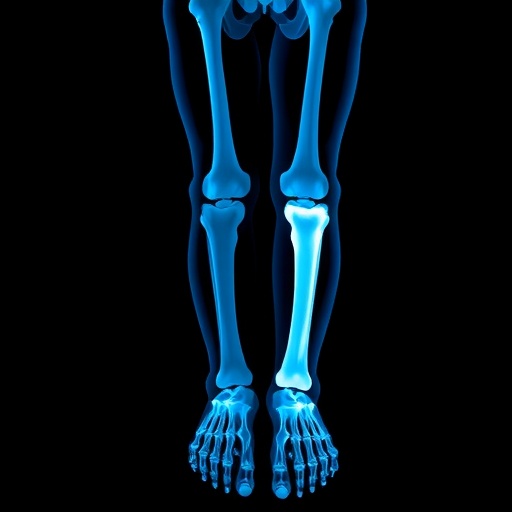
The methodological rigor of the study is particularly noteworthy. Employing advanced magnetic resonance imaging (MRI) combined with quantitative three-dimensional modeling, the researchers quantified muscle volume and bone length with remarkable precision. This approach minimized invasive procedures and radiation exposure, making repeated measurements feasible in a sensitive pediatric population. The combination of imaging data with robust statistical modeling allowed for discerning growth patterns across age groups ranging from early childhood into adolescence, ensuring a comprehensive understanding of time-dependent developmental shifts.
One of the cardinal findings reveals significant disparities in muscle growth trajectories between typically developing children and those with cerebral palsy. While muscle volume in typically developing children exhibits a predictable exponential increase during the critical growth phases, children with CP display attenuated muscle expansion, marked by both reduced volume and altered muscle architecture. This impairment was more pronounced in distal muscles of the lower leg, potentially exacerbating functional limitations related to walking and balance. Such revelations underscore the necessity for targeted therapeutic interventions focused on muscle hypertrophy in cerebral palsy management.
Simultaneously, the study brings to light intriguing patterns in bone growth. Contrary to initial hypotheses suggesting that bone elongation closely mirrors muscle development, the researchers found that lower leg bones in children with cerebral palsy, although shorter on average, follow a growth trajectory not entirely synchronized with muscle deficits. This decoupling implies that neurological impairments inherent to CP might uniquely disrupt mechanotransduction pathways that regulate bone adaptation, a hypothesis that merits further cellular and molecular investigation.
Interpreting these findings within the broader biomechanical context, the study emphasizes the intricate interdependence between muscle forces and skeletal form during growth. In healthy development, mechanical loading generated by muscle contraction promotes bone modeling, ensuring structural integrity and functional optimization. The attenuated muscle growth in children with CP likely results in diminished mechanical stimulation, altering bone quality and geometry subtly yet significantly. Such biomechanical insights are critical for devising physiotherapeutic regimens that stimulate muscle activation to indirectly benefit bone health.
Moreover, the study’s longitudinal nature allows for observing critical windows where intervention might yield maximal benefits. The researchers highlight an early childhood phase as a potentially optimal period for therapeutic engagement, owing to heightened plasticity in musculoskeletal tissues during rapid growth. Interventions leveraging resistance training, neuromuscular electrical stimulation, or pharmacological agents could be timed strategically to harness this window, maximizing gains in muscle strength and bone robustness.
The study also presents a nuanced discussion on ambulatory status among children with CP. By focusing solely on ambulant children, the research delineates the spectrum of musculoskeletal compromise within milder functional severity. This distinction is essential since ambulatory children maintain varying degrees of mobility, which influences mechanical loading patterns and therefore growth dynamics. Future investigations might expand to non-ambulant populations to delineate the full spectrum of CP-related musculoskeletal adaptations.
In addition, Bolsterlee and colleagues incorporate a sophisticated statistical framework that accounts for individual variability by integrating mixed-effects models. This approach not only enhances the robustness of the results but also underscores the personalized nature of musculoskeletal growth trajectories. Such data-driven personalization is pivotal in tailoring clinical interventions to the unique growth patterns of each child, moving toward precision rehabilitation.
The implications of this study resonate beyond clinical rehabilitation, extending into the realm of biomedical engineering and device development. Understanding detailed muscle and bone growth patterns can inform the design of orthotic devices, prosthetics, and robotics that adapt dynamically with the child’s development, offering improved fit, comfort, and efficacy. Given the rapid evolution of assistive technologies, integrating growth data enhances the translational impact of these innovations.
Furthermore, the authors call attention to the potential genetic and epigenetic regulators underlying the observed growth discrepancies. While this study did not directly assess molecular drivers, the phenotypic data lay the groundwork for interdisciplinary research combining imaging with genomic and proteomic profiling. Such integrative approaches could unravel the complex biological networks influencing musculoskeletal development in neurological conditions, ultimately guiding novel therapeutic targets.
Crucially, the research methodology highlights ethical and practical advances in pediatric studies. The non-invasive imaging protocol, coupled with a design minimizing participant burden through a mixed longitudinal approach, demonstrates an optimal balance between scientific rigor and participant welfare. This paradigm can serve as a benchmark for future longitudinal pediatric research in similarly vulnerable populations.
As emerging technologies such as AI-driven image analysis and biomechanical simulation mature, the potential to refine and automate muscle-bone growth assessments exponentially increases. This study exemplifies how integrating multidisciplinary tools pushes the envelope of pediatric developmental biology, enabling more precise and clinically actionable insights.
In sum, Bolsterlee, Chow, Davies, and their team’s seminal work offers a transformative window into the complex tapestry of musculoskeletal growth in children with and without cerebral palsy. By elucidating distinct growth patterns and their biomechanical underpinnings, the study paves the way for improved therapeutic strategies, personalized rehabilitation, and enhanced quality of life for affected children globally. This research not only advances fundamental biomedical knowledge but also exemplifies the power of longitudinal, data-rich investigations in pediatric science.
Looking ahead, the findings ignite a rich array of questions ripe for exploration. How might emerging regenerative therapies interface with impaired muscle-bone crosstalk in CP? Can nutritional interventions synergize with physical therapies to optimize growth outcomes? What role do neural plasticity and motor control exert during these critical developmental phases? Addressing these will invariably build upon the robust foundational knowledge this study provides.
Ultimately, this research stands as a beacon highlighting the profound complexity of human development when intersected by neurological disorders. Its blend of technical sophistication, clinical relevance, and forward-looking perspectives embodies the essence of transformative biomedical discovery. The pediatric community, from clinicians to researchers, can harness these insights to usher in a new era of integrative care tailored to the nuanced realities of childhood growth and development.
Subject of Research: Muscle and bone growth in the lower legs of typically developing children and ambulant children with cerebral palsy.
Article Title: Muscle and bone growth in the lower legs of typically developing children and ambulant children with cerebral palsy: a mixed longitudinal study.
Article References:
Bolsterlee, B., Chow, B.V.Y., Davies, S. et al. Muscle and bone growth in the lower legs of typically developing children and ambulant children with cerebral palsy: a mixed longitudinal study. Pediatr Res (2025). https://doi.org/10.1038/s41390-025-04558-0
Image Credits: AI Generated
DOI: 19 November 2025
Tags: biomechanics of muscle and bonebone growth in children with CPcerebral palsy muscle developmentfunctional independence in children with disabilitiesimaging techniques in growth studieslongitudinal study on CPlower leg growth in childrenmuscle volume measurement in childrenneurological conditions affecting growthpediatric musculoskeletal researchpediatric rehabilitation sciencequality of life in CP


