A retrospective radiological investigation has demonstrated that machine learning algorithms applied to routine non-contrast chest computed tomography scans can accurately identify abnormal bone mineral density in individuals diagnosed with type 2 diabetes mellitus. Reporting in Frontiers in Endocrinology, researchers from The Second Affiliated Hospital of Shandong First Medical University evaluated six distinct supervised learning architectures trained on high-throughput radiomic features extracted from the first lumbar vertebra. Among the evaluated algorithms, a linear support vector machine emerged as the most balanced and resilient framework, achieving an area under the receiver operating characteristic curve of 0.945 and an overall accuracy of 0.906 during nested cross-validation. By utilizing standard thoracic imaging datasets acquired for unrelated medical evaluations, this computational approach provides a viable mechanism for opportunistic skeletal health screening, identifying compromised bone mass without subjecting patients to supplemental radiation exposure, specialized bone densitometry hardware, or external calibration phantoms.
The clinical motivation for opportunistic screening stems directly from the complex systemic pathology linking type 2 diabetes mellitus with progressive skeletal degradation. As chronic metabolic dysregulation impairs bone turnover, individuals with diabetes exhibit altered bone micro-architecture and diminished mechanical strength, significantly elevating their susceptibility to fragility fractures. When osteoporotic fractures occur in diabetic populations, they frequently trigger severe secondary clinical sequelae, including permanent mobility impairment, accelerated renal decline, localized and systemic tissue infections, compromised bone healing, and increased patient mortality. These debilitating complications collectively impose substantial medical burdens and financial expenditures on healthcare infrastructures worldwide.
Conventional clinical assessment of bone mineral density relies predominantly on dual-energy X-ray absorptiometry, which currently serves as the established international reference standard for diagnosing osteoporosis. However, dual-energy X-ray absorptiometry has limitations as a screening method for osteoporosis, prompting a growing consensus to seek accurate alternative technologies. Because bone mineral density derived from dual-energy X-ray absorptiometry can be influenced by degeneration of spine disease and calcification of the abdominal aorta, it cannot accurately reflect real bone mineral density. Quantitative computed tomography overcomes these geometric limitations by delivering true volumetric, three-dimensional multi-planar visualizations unaffected by adjacent soft tissues, allowing radiologists to isolate and quantify cancellous trabecular bone separately from cortical compartments. Nevertheless, quantitative computed tomography mandates specialized analysis software and dedicated calibration phantoms that restrict its implementation during standard clinical visits, highlighting the necessity for opportunistic screening frameworks that leverage existing routine computed tomography scans.
To establish and evaluate the machine learning screening framework, the investigators retrospectively enrolled 249 consecutive patients with type 2 diabetes mellitus who underwent concurrent non-contrast-enhanced chest computed tomography and quantitative computed tomography examinations between January 2021 and June 2024. The study protocol was formally approved by the Institutional Ethics Committee on Biomedical Research under approval number 2024-H-040, with written informed consent waived due to its retrospective design in accordance with the Declaration of Helsinki. The study cohort ranged in age from 40 to 97 years, with a mean age of 60.93 ± 11.49 years. Eligibility required patients to be at least 40 years old, satisfy the World Health Organization diagnostic criteria established in 1999 for type 2 diabetes mellitus, and have undergone baseline thoracic imaging and laboratory testing within an interval of seven days or less.
Rigorous clinical and radiological exclusion criteria were systematically enforced to prevent confounding influences on bone metabolism and image quality. The researchers excluded individuals with concurrent endocrine disorders, hematological malignancies, severe end-stage renal disease, active liver dysfunction, or other malignant neoplasms. Patients who had received pharmacological agents known to alter bone metabolism within the six months preceding imaging were likewise omitted. Following these stringent screening procedures, the remaining 249 qualified patients were randomly partitioned using a 7:3 allocation ratio into a primary model development training cohort consisting of 175 patients and an entirely independent internal test cohort comprising 74 patients reserved solely for downstream validation.
Baseline demographic and clinical profiling of the enrolled cohort revealed that abnormal bone mineral density was present in 40.56 percent of the participants, representing 101 out of the 249 total patients, distributed between 52 men and 49 women. The remaining 59.44 percent, comprising 148 individuals including 32 men and 116 women, exhibited normal skeletal density. When comparing patient subsets, those with abnormal bone density were significantly older than their counterparts with preserved bone mass, recording a mean age of 68.00 ± 10.61 years compared to 56.10 ± 9.39 years. Interestingly, circulating glycated hemoglobin concentrations did not differ significantly between normal and abnormal bone density groups. Quantitative computed tomography diagnostic criteria categorized patients based on average volumetric density measured across the twelfth thoracic and first lumbar vertebral bodies, classifying values exceeding 120 milligrams per cubic centimeter as normal, values between 80 and 120 milligrams per cubic centimeter as low bone mass, and measurements beneath 80 milligrams per cubic centimeter as osteoporosis.
All thoracic computed tomography examinations were acquired on a single 256-slice scanner utilizing a uniform technical protocol consisting of a 120 kilovolt-peak tube voltage, automatic tube current modulation, and reconstructed slice thickness of 1.25 millimeters using a standard bone reconstruction kernel. Intravenous contrast media were deliberately omitted to prevent artificial fluctuations in Hounsfield units caused by variable vascular and parenchymal enhancement. To extract phenotypic bone characteristics, two independent radiologists with a decade of specialized clinical experience performed semi-automatic three-dimensional volume-of-interest segmentations of the cancellous bone inside the first lumbar vertebral body using 3D Slicer software. Both readers operated under strict blinding regarding patient clinical records and diagnostic classifications, resolving boundary discrepancies through consensus. Standardized image preprocessing was uniformly applied across the segmented volumes, encompassing isotropic voxel resampling to one cubic millimeter via B-spline interpolation, gray-level discretization using a fixed bin width of 25 Hounsfield units, and zero-mean unit-variance distribution normalization to eliminate acquisition heterogeneity.
From the segmented cancellous volumes of interest, the automated Research Portal workstation extracted 2273 high-dimensional radiomic features capturing volumetric shapes, localized intensity variations, and subtle micro-architectural texture patterns. To prevent dimensionality-induced model overfitting, the investigators applied the minimum redundancy-maximum relevance feature selection algorithm across the 175-patient training set, systematically benchmarking candidate feature subsets containing 5, 10, 15, and 20 attributes. The 15-feature subset attained the highest mean area under the curve of 0.932, outperforming the candidate sets of 5 features at 0.920, 10 features at 0.922, and 20 features at 0.927. Consequently, these 15 optimal radiomic signatures were selected to configure six supervised machine learning models: Random Forest, Support Vector Machines with Radial Basis Function kernel, Support Vector Machines with Linear kernel, Neural Network, Extreme Gradient Boosting Linear, and K-Nearest Neighbors.
Internal model tuning and validation were conducted exclusively within the training set using a nested cross-validation architecture to mitigate algorithmic optimism. The outer validation loop executed 100 repetitions of leave-group-out cross-validation, while inner five-fold cross-validation managed hyperparameter optimization across all algorithms while maintaining the 15 selected radiomic features fixed. Across these 100 iterations, the linear support vector machine achieved the leading diagnostic efficacy, exhibiting a mean accuracy of 0.906 ± 0.032, an area under the curve of 0.945 ± 0.028, sensitivity of 0.896 ± 0.070, specificity of 0.911 ± 0.058, positive predictive value of 0.881 ± 0.071, and negative predictive value of 0.931 ± 0.042. The neural network model performed comparably, recording an accuracy of 0.906 ± 0.031 and an area under the curve of 0.941 ± 0.029, with no statistically significant differences observed between these two top architectures.
Algorithmic stability was quantitatively evaluated by calculating the relative standard deviation across the 100 leave-group-out iterations to determine resilience against data perturbations. While the neural network exhibited the lowest relative standard deviation for mean accuracy at 3.40 percent compared to 3.54 percent for the linear support vector machine, the linear support vector machine demonstrated superior consistency in discriminative power, producing the lowest relative standard deviation for mean area under the curve at 3.04 percent versus 3.08 percent for the neural network. In contrast, remaining architectures demonstrated reduced stability or lower discriminatory capacity: Random Forest produced an area under the curve of 0.937 with a relative standard deviation of 3.18 percent; Extreme Gradient Boosting achieved an area under the curve of 0.929 with 3.41 percent deviation; radial support vector machines showed an accuracy of 0.896 with 3.76 percent deviation; and K-Nearest Neighbors yielded the lowest area under the curve at 0.925 with 3.81 percent deviation.
When challenged with the unseen 74-patient internal test set, the frozen linear support vector machine verified its diagnostic reliability, securing an area under the curve of 0.913 with a 95 percent confidence interval spanning 0.834 to 0.993. The algorithm achieved an overall classification accuracy of 0.892, accompanied by a specificity of 0.955, positive predictive value of 0.923, negative predictive value of 0.875, and sensitivity of 0.800. Minor calibration deviations occurred solely at the extreme boundaries of the risk spectrum, which the authors attributed to the modest size of the test cohort. Furthermore, decision curve analysis established positive net clinical benefit across a broad spectrum of decision thresholds, verifying practical clinical utility.
The study authors highlighted several methodological limitations that warrant careful consideration before clinical implementation. First, the investigation was structured as a single-center retrospective study with a relatively small sample size of 249 patients, underscoring the necessity for prospective, large-scale multicenter trials to establish external generalizability across diverse scanners and populations. Second, the model was validated solely through internal held-out test data rather than an independent external hospital cohort. Third, radiomic features were extracted solely from three-dimensional segmentation of cancellous bone, which might not completely capture alterations within the bone microenvironment. Finally, the model was optimized to identify abnormal volumetric bone density thresholds defined by quantitative computed tomography rather than predicting incident fracture events directly, meaning the model serves as an auxiliary opportunistic screening instrument rather than an absolute predictor of bone fragility.
Subject of Research: Machine learning models for opportunistic screening of abnormal bone mineral density in type 2 diabetes using routine chest CT radiomics
Article Title: Opportunistic application of machine learning for bone health stratification in type 2 diabetes mellitus
Article References: Yang, H., Chen, X., Jia, C., Yao, Q., & Liu, M. (2026). Opportunistic application of machine learning for bone health stratification in type 2 diabetes mellitus. Frontiers in Endocrinology, 17, Article 1935105. https://doi.org/10.3389/fendo.2026.1935105
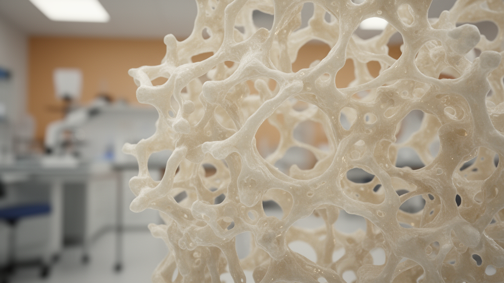
Image Credits: AI Generated
DOI: 10.3389/fendo.2026.1935105
Keywords: type 2 diabetes mellitus, bone mineral density, opportunistic screening, chest computed tomography, radiomics, linear support vector machine, quantitative computed tomography, osteoporosis, first lumbar vertebra, cancellous bone, machine learning, feature extraction, minimum redundancy maximum relevance, nested cross validation, bone health, medical imaging, trabecular bone
News Source: Ophelia Keating. (November 11, 2026). Linear Support Vector Machine Accurately Identifies Abnormal Bone Density in Diabetic Patients. Scienmag.