A groundbreaking advancement in the visualization of molecular chirality promises to reshape the way scientists study the “handedness” of molecules and materials. Chirality, a fundamental property describing how certain structures exist in left- or right-handed forms, plays a critical role in everything from the scent of a mint leaf to the effectiveness of life-saving medicines. Researchers at ETH Zurich, led by Professor Romain Quidant, have pioneered an innovative imaging method that captures chirality in unprecedented spatial detail using a single image, significantly surpassing the traditional approach of measuring chirality as an averaged property across a sample.
Understanding chirality is essential because molecules that are nearly identical in composition can behave drastically differently based on their three-dimensional orientation. This phenomenon is evident in daily life — for instance, caraway seeds and spearmint contain molecules with nearly identical structures but emit very different aromas because they are mirror images, or enantiomers, of each other. Similarly, many pharmaceuticals owe their efficacy to this molecular asymmetry, as one enantiomer of a drug molecule may be therapeutic while its mirror image is ineffective or even toxic. Until now, studying these variations at a detailed, localized scale has posed a formidable challenge.
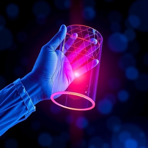
The team’s novel technique leverages the interaction between chiral samples and circularly polarized light to generate a spatial map of handedness within a sample. Circular polarization refers to light waves whose electric field vectors rotate in a spiral as the light propagates, either clockwise or counterclockwise. Chiral molecules and nanostructures interact differently with these two types of circularly polarized light, absorbing or rotating the light’s oscillations in distinct ways depending on their handedness. Capitalizing on these optical signatures, the researchers developed a single-shot wide-field spectroscopic imaging method to spatially resolve chirality.
.adsslot_lBqXC4yeKY{width:728px !important;height:90px !important;}
@media(max-width:1199px){ .adsslot_lBqXC4yeKY{width:468px !important;height:60px !important;}
}
@media(max-width:767px){ .adsslot_lBqXC4yeKY{width:320px !important;height:50px !important;}
}
ADVERTISEMENT
What sets this approach apart from traditional methods is its ability to capture both left- and right-circularly polarized light interactions simultaneously in one image, rather than requiring separate measurements for each polarization state. To accomplish this, the team crafted a sophisticated optical system that splits the light transmitted through the sample into its circularly polarized components by employing carefully engineered reference beams. These beams create interference patterns that encode how the sample differentially interacts with left- and right-handed circular polarizations. While these overlapping patterns are incomprehensible to the naked eye, computational algorithms decode the data to produce clear, color-coded maps of the chiral distribution across the sample surface.
In a laboratory demonstration, the researchers applied their imaging technology to artificial nanostructures fabricated from gold, which were deliberately designed with varying handedness. The experiment validated the method’s ability to accurately distinguish left-handed from right-handed regions, visualizing intricate shapes such as letters “L” and “R” composed of nanoscale chiral elements. This ability to spatially resolve chirality at the nanoscale is a marked improvement over existing techniques, which only yield average information and cannot reveal heterogeneity in the sample’s handedness.
The implications of this breakthrough extend well beyond physics and nanotechnology, offering transformative potential for diverse fields such as biology, medicine, and materials science. Biological tissues and cells often exhibit chirality not only at the molecular level but also in larger structural arrangements. However, characterizing these spatial variations without damaging the sample or relying on chemical staining has been an elusive goal. The imaging method pioneered by Quidant’s group could offer a non-invasive way to detect and map chirality differences between healthy and diseased tissues, potentially leading to novel diagnostic tools.
In the realm of material engineering, many advanced materials display spatially varied chirality, which influences their optical, mechanical, and chemical properties. Until now, this spatial variation has been difficult to assess directly. The new technique provides researchers with a powerful tool to visualize handedness patterns within complex materials, enabling deeper insight into how chirality affects macroscopic behavior and facilitating the design of new materials with tailored chiral features.
Pharmaceutical sciences stand to benefit significantly from this technology as well. Since many drugs are chiral, with only one enantiomer providing the desired therapeutic effect, being able to map the chirality distribution within complex mixtures or formulations can improve drug design, quality control, and analytical procedures. This imaging approach may enable chemists and pharmacologists to identify chiral impurities or quantify enantiomeric excess with spatial specificity, thereby refining drug safety and efficacy.
Despite its promise, the technique remains at an early stage of development. One of the main challenges the team has faced is the intrinsic weakness and noise sensitivity of the optical signals associated with chirality at the nanoscale. To ensure that the measured signals genuinely stem from chiral interactions rather than artefacts, the researchers implemented meticulous noise-reduction strategies and signal processing protocols. Future work will focus on enhancing the system’s sensitivity and robustness to enable practical applications outside the laboratory environment.
Moving forward, the researchers are keen to collaborate with experts across various disciplines to explore potential use cases and optimize the system for real-world scenarios. As lead author Rebecca Büchner notes, while the platform’s capabilities are well understood by the team, other scientists might identify novel applications and tailor the technology to address domain-specific questions. Whether in clinical diagnostics, material characterization, or pharmaceutical analysis, the ability to map chirality spatially with a single image heralds a new era of chiral research.
Ultimately, this pioneering work illustrates the power of merging advanced optics, nanotechnology, and computational imaging to solve longstanding scientific challenges. By making the invisible spatial patterns of molecular handedness visible, it opens the door to a deeper understanding of chiral phenomena that underpin chemistry, biology, and materials science. The pathway from this fundamental research to transformative applications may be long, but the potential impact on science and technology is immense.
This research was recently published in the prestigious journal Nature Photonics, highlighting the interdisciplinary collaboration and technical ingenuity underpinning the advance. With continued development and collaboration, spatially resolved chirality imaging could soon become a staple in laboratories worldwide, enriching our ability to see and study the asymmetrical world at the heart of matter.
Subject of Research: Imaging and visualization of molecular and nanoscale chirality through wide-field spectroscopic methods.
Article Title: Wide-field spectroscopic imaging of optical activity
News Publication Date: 28-Jul-2025
Web References: https://doi.org/10.1038/s41566-025-01722-0
Keywords: chirality, handedness, circularly polarized light, nanophotonics, optical activity, nanostructures, spectroscopy, spatial imaging, molecular asymmetry, drug efficacy, biomedical imaging
Tags: challenges in chirality measurementchirality’s impact on scent and flavorenantiomers in pharmaceuticalsETH Zurich research advancementsinnovative imaging methods in chemistryleft-handed and right-handed moleculesmolecular chirality visualizationnanostructures imaging techniquesProfessor Romain Quidant’s researchsignificance of molecular handedness in sciencespatial detail in chirality studiesthree-dimensional molecular orientation


