
Cell biology has made significant strides in recent years, especially with regard to imaging and segmentation techniques that enhance our understanding of microscopic structures. Among the most notable advancements is the introduction of Cellpose3, a groundbreaking tool designed for the restoration and segmentation of distorted microscopy images. This innovative algorithm is revolutionizing the field by allowing scientists to effectively identify and delineate individual cells, even in challenging conditions, where noise, blurring, or undersampling might have previously hindered their observations.
The third iteration of Cellpose, known as Cellpose3, addresses a critical issue faced by researchers: the inability to accurately discern cellular boundaries within images that do not meet optimal quality standards. Traditional methods for segmenting cell images often fall short in scenarios where artifacts distort the visual data, which can produce unreliable results. The developers, Carsen Stringer and Marius Pachitariu from the Janelia Group, recognized the need for a more robust solution and set out to create an adaptive algorithm that caters specifically to such adversities. By training Cellpose3 to enhance segmentation rather than merely improving image quality, they brought a transformative approach to the scientific community.
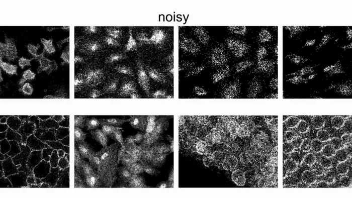
Cellpose3 operates through a sophisticated restoration algorithm that converts blurred and noisy images into clearer, more defined representations. This capability not only facilitates precise segmentation by the original Cellpose algorithm but also streamlines the workflow for researchers. Instead of spending countless hours manually adjusting and refining images, users can apply the restoration algorithm with a simple click of a button integrated into the Cellpose application. This user-friendly approach ensures that researchers can focus their efforts on analysis and interpretation, rather than being bogged down in technical preprocessing.
In a bid to enhance the versatility of Cellpose3, the algorithm was trained on an extensive and diverse collection of images. This training set comprises various microscopy scenarios, enabling the algorithm to generalize across different types of data. Consequently, it empowers users to deploy Cellpose3 effectively on their unique datasets without the need for extensive customization or prior experience in image processing techniques. As a result, this democratization of technology paves the way for broader applications across disciplines in life sciences.
The implications of such advancements extend far beyond mere convenience. Accurate cellular segmentation is essential for a wide range of biological inquiries, from studying cellular morphology and proliferation to understanding complex interactions within tissues. Cellpose3’s enhanced capabilities will enable researchers to uncover new insights into cellular dynamics, potentially ushering in breakthroughs in fields as diverse as cancer research, developmental biology, and neurobiology. By facilitating the study of cellular structures more effectively, Cellpose3 serves as a powerful asset in the ongoing mission to decode life’s complexities.
Another notable aspect of Cellpose3’s design is its conscientious integration of machine learning techniques. The algorithm’s training mechanism adeptly learns from the features present in images, capturing intricate details that are often overlooked by human analysts. Through leveraging deep learning methodologies, the algorithm continually refines its approach, resulting in improved performance over time. As new data is collected, Cellpose3 can adapt and enhance its predictive capabilities, making it an ever-improving tool in the hands of researchers.
Moreover, the availability of Cellpose3 in a community-driven application reinforces its potential. As researchers share their experiences and datasets, they contribute to the collective knowledge base that feeds back into the algorithm’s development. This iterative process of improvement embodies the spirit of collaboration and innovation that defines modern scientific endeavors. As more researchers adopt Cellpose3, it not only signifies a shift in microscopy practices but also fosters a culture of openness and shared progress in the scientific community.
The accessibility of such sophisticated tools can significantly alter the landscape of microscopy in both academic and industrial settings. By providing researchers with the ability to restore and segment images with an unprecedented level of ease and reliability, Cellpose3 empowers them to tackle complex biological questions that were previously out of reach. Researchers are now equipped to address pressing questions about cellular interactions, the effects of pharmacological agents, and the mechanisms of disease pathology, creating a ripple effect across numerous areas of study.
Looking ahead, the impact of tools like Cellpose3 on future scientific explorations cannot be overstated. As cellular imaging becomes increasingly vital in various realms of biomedicine and biotechnology, the need for accurate segmentation tools will only grow. In this context, Cellpose3 stands at the forefront of innovation, ready to meet the demands of modern research agendas and facilitate discoveries that can change our understanding of life at the cellular level.
Notably, the tool arises at a pivotal moment when rapid advancements in microscopy techniques, such as super-resolution imaging, are becoming mainstream. As these high-resolution strategies become more prevalent, the ability to process and analyze the resulting data efficiently is paramount. Cellpose3 not only complements these advancements but also empowers researchers to leverage the full potential of ultra-high-resolution microscopy by ensuring that the segmentation process is not a limiting factor.
In conclusion, as Cellpose3 continues to gain traction within the scientific community, it represents a significant milestone toward harnessing the true power of microscopy. Its adaptability and user-friendly application promise to revolutionize how scientists approach cellular imaging, presenting an exciting avenue for exploration and discovery. Researchers can look forward to an era where understanding the fundamental processes of life becomes more accessible and efficient, ultimately contributing to the advancement of science and medicine.
The future of tools like Cellpose3 is undoubtedly bright, with the potential to inspire new research directions and methodologies. As the community embraces these advanced technologies, we remain optimistic about the discoveries that lie ahead.
Subject of Research: Cellpose3 Algorithm for Image Restoration and Segmentation
Article Title: Cellpose3: One-Click Image Restoration for Improved Cellular Segmentation
News Publication Date: 12-Feb-2025
Web References: Cellpose, Research Article
References: Stringer, C., & Pachitariu, M. (2025). Cellpose3: one-click image restoration for improved cellular segmentation. Nature Methods. DOI: 10.1038/s41592-025-02595-5
Image Credits: Credit: Stringer and Pachitariu
Keywords
Image processing, segmentation techniques, microscopy, cell biology, automation in research, machine learning applications.
Tags: adaptive algorithms for cell biologyadvanced imaging techniquesalgorithmic approaches to cell identificationcell boundary detectionCellpose3challenges in cell imagingimage quality enhancement in microscopyinnovative solutions for microscopymicroscopy image segmentationnoise reduction in microscopyrestoration of distorted imagesscientific advancements in cell biology


