In a groundbreaking development that promises to revolutionize the way we study engineered heart tissues, researchers have unveiled a novel imaging technique harnessing the power of label-free mid-infrared dichroism-sensitive photoacoustic microscopy. This inventive approach enables unprecedented histostructural analysis of cardiac tissues without the need for exogenous dyes or markers, promising to enhance the precision and depth of cardiac tissue characterization.
At the heart of this innovation is the combination of mid-infrared light, known for its ability to probe molecular vibrations, with photoacoustic microscopy, a technique that converts absorbed optical energy into acoustic signals. The integration of dichroism sensitivity adds a new dimension—it allows the detection of anisotropic molecular orientations within tissue samples. This is particularly significant in the context of engineered heart tissues, where the alignment and structural organization of cellular and extracellular matrix components critically dictate tissue function.
Histological analysis has long been essential for evaluating tissue architecture during the development and maturation of bioengineered cardiac constructs. Traditional staining and imaging methods, while valuable, often require invasive preparation steps that can alter tissue integrity and are limited in their ability to offer molecular-level insights. The newly developed mid-infrared dichroism-sensitive photoacoustic microscopy technique addresses these limitations by providing a label-free methodology that preserves native tissue state, enabling high-resolution, chemically specific imaging.
The principle of operation hinges on the unique absorption properties of molecular bonds in the mid-infrared spectral region. As mid-infrared light excites vibrational modes of molecules such as proteins and lipids, the subsequent non-radiative relaxation generates localized thermoelastic expansion. This produces acoustic waves detectable by ultrasound transducers, forming the basis of photoacoustic microscopy. By exploiting dichroism—the dependency of absorption on the polarization direction of incident light—the system reveals anisotropic molecular arrangements, unveiling detailed structural alignment within the tissue.
Engineered heart tissues require meticulous structural organization to emulate native myocardial function, which is primarily governed by the orientation of contractile proteins and extracellular matrix fibers. The capacity of this technology to detect and image such molecular anisotropy noninvasively not only accelerates tissue characterization but also opens avenues for real-time monitoring of tissue maturation during cultivation.
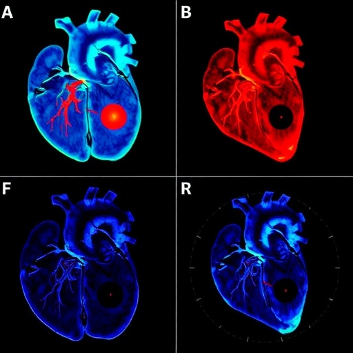
The researchers demonstrated this method on various engineered cardiac constructs, highlighting its capability to visualize intricate histological features such as fiber alignment, cell distribution, and extracellular matrix composition. Compared to conventional imaging modalities, the label-free mid-infrared photoacoustic approach offers superior chemical specificity without compromising spatial resolution—facilitating direct correlation between structural features and functional properties of the tissue.
Moreover, the utilization of mid-infrared wavelengths addresses a critical challenge in biomedical imaging: the trade-off between penetration depth and molecular specificity. While shorter wavelengths enable higher resolution, they lack chemical contrast, and longer wavelengths often suffer from limited tissue penetration. The photoacoustic effect circumvents these issues by detecting ultrasound signals rather than light directly, enabling the deep interrogation of thick tissue samples without sacrificing molecular detail.
Another advantage of this platform is its compatibility with live tissue environments, potentially allowing longitudinal studies of tissue development and disease progression. This dynamic monitoring capability is transformative for regenerative medicine, where the functionality of bioengineered tissues must be validated prior to transplantation or therapeutic use.
The detailed spectroscopic information afforded by the system enhances the diagnostic potential beyond structural imaging. By discerning specific molecular fingerprints, it could help identify pathological changes or deviations in tissue composition indicative of disease states or insufficient tissue engineering protocols. This diagnostic precision paves the way for personalized medicine applications, where tailored treatments depend on an accurate understanding of tissue microenvironments.
Integration of this photoacoustic microscopy paradigm with existing cardiac tissue engineering workflows promises to streamline the validation process of tissue constructs. Researchers and clinicians can benefit from expedited, non-destructive assessments that preserve valuable samples for further analysis or therapeutic application. This approach fosters a more efficient pipeline from laboratory development to clinical translation.
The mid-infrared dichroism-sensitive photoacoustic microscopy embodies a convergence of optical physics, acoustics, and bioengineering, showcasing how interdisciplinary strategies can surmount longstanding challenges in biomedical imaging. As the technology matures, scaling and automation may facilitate its adoption in routine tissue analysis laboratories and regenerative medicine clinics worldwide.
Future directions may include expanding the approach to other tissue types where molecular orientation and composition critically influence function, such as neural, musculoskeletal, and connective tissues. Additionally, coupling with machine learning algorithms for image analysis could accelerate data interpretation, enabling rapid phenotyping and quality control of engineered tissues at scale.
The implications of this innovation extend beyond engineered heart tissues alone. The foundational principles could spur a new generation of label-free imaging techniques that capture the complexities of tissue biology with minimal preparation and maximal informational content. This shift toward non-invasive, chemically informative imaging heralds a new era in histopathology and tissue engineering research.
By enabling detailed visualization of histostructural features, this mid-infrared photoacoustic microscopy method holds the promise to deepen our understanding of cardiac biology and enhance the development of therapies for heart disease. As cardiovascular conditions remain a leading cause of morbidity and mortality globally, tools that refine engineered tissue characterization are vital for advancing regenerative solutions.
In summary, the advent of label-free mid-infrared dichroism-sensitive photoacoustic microscopy marks a significant leap forward in the field of biomedical imaging and tissue engineering. Its ability to combine chemical specificity, structural resolution, and deep tissue penetration without the need for exogenous labels positions it as an indispensable tool for the future of cardiac tissue research and therapy development.
As this technique gains wider acceptance and technical refinements, it is poised to become a cornerstone method for the histostructural analysis of engineered tissues, ultimately contributing to improved clinical outcomes for patients suffering from heart disease and possibly a broad spectrum of other disorders where tissue architecture is a critical parameter.
Subject of Research: Histostructural analysis of engineered heart tissues using label-free mid-infrared dichroism-sensitive photoacoustic microscopy.
Article Title: Label-free mid-infrared dichroism-sensitive photoacoustic microscopy for histostructural analysis of engineered heart tissues.
Article References:
Park, E., Hwang, D.G., Choi, H. et al. Label-free mid-infrared dichroism-sensitive photoacoustic microscopy for histostructural analysis of engineered heart tissues. Light Sci Appl 15, 49 (2026). https://doi.org/10.1038/s41377-025-02117-0
Image Credits: AI Generated
DOI: 10.1038/s41377-025-02117-0
Tags: advanced histological techniquesanisotropic molecular orientation detectionbioengineered cardiac constructscardiac tissue characterizationcardiac tissue integrity preservationengineered heart tissues analysishistostructural analysis methodslabel-free imaging techniquesmid-infrared photoacoustic microscopymolecular vibrations imagingnon-invasive tissue imagingphotoacoustic imaging innovations


