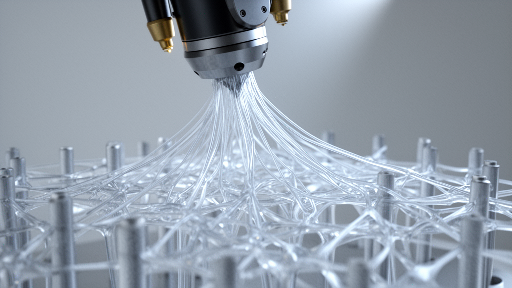
Researchers have developed a kinematically controlled manufacturing workflow that repurposes fused deposition modeling stringing from an uncontrolled printing defect into a reproducible method for producing suspended polymer microfibers. Reporting in Frontiers in Materials, an interdisciplinary team from the University Hospital of Würzburg, the University of Bayreuth, and the University Hospital Düsseldorf demonstrated that molten poly(lactic acid) can be drawn across open gaps into highly aligned fibrous architectures with microscale precision. In conventional material extrusion, unwanted filament oozing during non-extrusion travel movements—widely known as stringing—is treated as an operational failure that ruins part surface quality. Rather than suppressing this fluid dynamic behavior, the investigators systematically parameterized printer kinematics, thermal stability, and nozzle dynamics to convert stringing into a controllable drawing process. Synchronizing printhead acceleration, travel speed, and localized anchoring positions reliably produces free-spanning microfibers measuring approximately forty micrometers in diameter, creating standardized anisotropic microenvironments for three-dimensional tissue engineering without specialized microspinning equipment.
The development addresses a persistent challenge in cell biology, where conventional two-dimensional substrates fail to capture the complex mechanical and structural cues of living tissues. Monolayers cultured on flat surfaces lack the multiaxial mechanical constraints, biochemical gradients, and organized fibrillar architecture present in vivo, altering cellular morphology, signaling dynamics, and gene expression. Native musculoskeletal, cardiac, and neural tissues feature intrinsically anisotropic extracellular matrix fibers that guide directional migration, cellular alignment, and tissue maturation. Recreating this organization has remained difficult; while biological hydrogels provide three-dimensional environments, they often suffer from batch variability and non-standardized preparation. Conversely, electrospinning and near-field variants can produce aligned fiber meshes but struggle with spatial repeatability, electrostatic charge accumulation, and construct height limitations. Melt electrowriting and microfluidic spinning face related constraints regarding suspended fiber placement across large spans. By establishing a digitally defined additive manufacturing process capable of producing freestanding anisotropic microfiber arrays, the authors provide a reproducible platform for standardized tissue modeling.
To transform stringing into an engineered fabrication technique, the researchers upgraded the mechanical and thermal subsystems of a Voron 0.1 CoreXY desktop printer. High-speed drawing requires structural rigidity to suppress dynamic vibration during rapid directional changes and high accelerations. The team replaced critical frame elements and printhead mounts with CNC-milled aluminum components, stiffening the mechanical assembly. Material dispensing relied on a lightweight direct-drive Bondtech LGX-Lite extruder paired with a Mosquito Magnum hotend and a 0.3-millimeter pointed steel-tip nozzle for 1.75-millimeter filament. Because consistent fiber formation demands extreme thermal stability, boron nitride thermal paste was applied between heating elements and the hotend block, limiting nozzle temperature fluctuations to within plus or minus 0.3 degrees Celsius during fabrication. Motion was controlled by Klipper firmware running on a Raspberry Pi single-board computer connected to an SKR Mini E3 mainboard, enabling custom acceleration and velocity parameters necessary for rapid non-extrusion drawing between anchor points.
The printing strategy bypassed commercial slicing software in favor of mathematically defined toolpaths created in Python using the FullControl framework. Standard slicers automate layer generation but suppress travel stringing through retraction and path wiping, precluding intentional microfiber drawing. In contrast, the customized Python script directly encoded geometrical parameters, dispensing commands, dwell times, and non-extrusion travel vectors. The script first synthesized a rigid polygonal outer frame that provided anchoring perimeters for the suspended fibers. Individual fibers were formed by coordinating discrete polymer deposition at defined perimeter anchor coordinates with rapid non-extrusion movements bridging the frame. Crucially, the toolpath directed non-printing transit movements around the external boundary of the construct rather than across the central opening. This perimeter routing eliminated physical contact and convective heat exposure that could disrupt previously drawn, suspended microfibers, allowing multi-layered networks to be assembled without damaging delicate pre-existing spans.
Experimental kinematic characterization confirmed that the modified printer reached its required motion envelope while defining the physical process window governing fiber drawing. Using an MPU-6050 accelerometer logging at approximately forty-two samples per second, the researchers recorded peak accelerations of plus or minus twenty meters per second squared along the primary movement axis during active drawing. Kinematic calculations illustrated that printhead velocity depends directly on acceleration and available travel distance. For an available travel distance of 8.65 millimeters at twenty meters per second squared, the calculated theoretical maximum velocity was 0.59 meters per second. However, the authors selected an operating velocity of 0.33 meters per second, equivalent to twenty thousand millimeters per minute, which required only 2.7 millimeters of linear travel to attain. Operating within this submaximal velocity regime established a stable kinematic margin, reducing susceptibility to transient melt variations and mechanical fluctuations while ensuring continuous suspended fiber formation across the open frame.
Under these parameterized kinematic conditions, the system reproducibly generated suspended poly(lactic acid) microfibers with an average diameter of 39.8 plus or minus 3.5 micrometers. Stable fiber generation was achieved at a nozzle temperature of 180 degrees Celsius, a bed temperature of 60 degrees Celsius, and an initial anchoring extrusion length of two millimeters. Implementing a short dwell time of one hundred to three hundred milliseconds prior to printhead motion improved fiber continuity by allowing nozzle melt pressure and temperature to equilibrate. Morphological analysis revealed distinct structural features reflecting printhead kinematics: drawn fibers displayed tapered initiation regions of four hundred to six hundred micrometers before stabilizing at steady-state dimensions, while termination zones formed triangular or trapezoidal footprints of approximately five hundred micrometers upon reaching the opposing anchor. Lower temperatures or higher drawing velocities led to fiber thinning and breakages, whereas slower speeds or excess extrusion caused fiber thickening and unintended fusion.
The scripted workflow enabled the fabrication of complex multilayer architectures by stacking suspended fiber arrays with controlled spatial orientations. The researchers constructed three-layer hexagonal models featuring a lateral fiber spacing of 0.5 millimeters and an interlayer vertical spacing of three hundred micrometers, elevated 0.6 millimeters above the build plate on printed base layers. Successive layers were rotated sixty degrees, producing a regular triangular micropattern across the central aperture. Quantitative image analysis demonstrated high within-construct geometric consistency, yielding spacing coefficients of variation of 2.7, 4.4, and 4.1 percent for horizontal and diagonal fiber sets. Fiber orientation standard deviations remained below 0.25 degrees, with mean inter-layer angles measuring 60.77, 60.03, and 59.20 degrees, matching design targets. The team also demonstrated vertical scalability by printing fifteen-layer hexagonal constructs consisting of five three-layer units separated by solid spacing bands, achieving a total height of 5.55 millimeters while preserving open, freestanding microfiber geometry.
To verify mechanical integrity, individual suspended microfibers were subjected to uniaxial tensile testing. Twenty single fibers with a gauge length of seventeen millimeters were isolated and pulled to failure at a rate of 1.5 millimeters per minute under controlled ambient conditions. Stress calculations based on the reference mean fiber diameter demonstrated consistent linear-elastic behavior across all samples up to approximately one percent strain and twenty megapascals. The microfibers exhibited an average Young’s modulus of 2.3 plus or minus 0.3 gigapascals and an ultimate tensile strength of 32.4 plus or minus 6.6 megapascals. Elongation at break averaged 3.0 plus or minus 0.6 percent, with failure strains falling between 2.2 and 4.1 percent, yielding an average material toughness of 725 plus or minus 272 kilojoules per cubic meter. Tensile fracture occurred within the central homogeneous fiber spans rather than at anchor points or transition zones, confirming that drawing did not introduce structural vulnerabilities at anchoring interfaces.
Because untreated poly(lactic acid) lacks cell-adhesive motifs, the researchers evaluated post-fabrication chemical activation and biofunctional coating. Constructs underwent alkaline surface activation by incubation in 0.01 molar sodium hydroxide for twenty minutes, followed by washing with phosphate-buffered saline until reaching neutral pH. Samples were then coated with human fibronectin at ten micrograms per milliliter for twenty-four hours at thirty-seven degrees Celsius. Attenuated total reflectance Fourier-transform infrared spectroscopy confirmed retention of the polyester backbone across all treatments, displaying characteristic ester carbonyl peaks at 1751 reciprocal centimeters and carbon-oxygen stretching bands between 1000 and 1300 reciprocal centimeters. While fibronectin adsorption remained below the detection limit of infrared spectroscopy due to dominant substrate signals, sodium hydroxide-treated fibers displayed spectral features consistent with partial surface hydrolysis and formation of hydrophilic groups. These surface modifications created an amenable interface for anchorage-dependent cell attachment and subsequent three-dimensional biological culture.
Biological compatibility was evaluated by culturing mouse fibroblasts and motoneuron-like cells on the suspended scaffolds. Murine BALB/3T3 fibroblasts cultured for five days showed sparse, discontinuous attachment on untreated fibers, whereas alkaline-activated fibers promoted extensive cellular alignment along the fiber axes. Fibronectin coating produced the highest overall coverage and actin signal intensity, forming multi-layered cellular networks across adjacent fibers. When mouse motoneuron-like NSC-34 cells were embedded in Matrigel and cultured for three days, PrestoBlue metabolic assays confirmed cell viability and proliferation across all conditions. While relative proliferation fold change showed no statistically significant differences between groups, alkaline activation yielded significantly higher absolute signal at day three. Fluorescence imaging showed that fibronectin-coated scaffolds supported the densest three-dimensional cellular distribution, with motoneurons populating both individual fibers and inter-layer spaces. These findings demonstrate that kinematically controlled FDM stringing provides an accessible, reproducible platform for fabricating standardized anisotropic microenvironments for mechanobiology and tissue engineering.
Subject of Research: Kinematically controlled fused deposition modeling of suspended anisotropic microfiber scaffolds for tissue engineering
Article Title: Kinematically controlled FDM stringing of suspended anisotropic microfiber scaffolds for standardized 3D tissue engineering
Article References: Cortopassi, S., Schönhammer, P., Murenu, N., Lamberger, Z., Ryma, M., Zwingelberg, S., Schaefer, N., & Lang, G. (2026). Kinematically controlled FDM stringing of suspended anisotropic microfiber scaffolds for standardized 3D tissue engineering. Frontiers in Materials, 13, Article 1936349. https://doi.org/10.3389/fmats.2026.1936349
Image Credits: AI Generated
DOI: 10.3389/fmats.2026.1936349
Keywords: fused deposition modeling, polymer stringing, suspended microfibers, poly(lactic acid), anisotropic scaffolds, 3D cell culture, CoreXY 3D printer, motion kinematics, FullControl toolpaths, tensile strength, surface activation, fibronectin coating, BALB/3T3 fibroblasts, NSC-34 motoneurons, mechanobiology, biofabrication, tissue engineering
News Source: Denise Maddox. (November 18, 2026). Kinematically Controlled 3D Printing Transforms Polymer Stringing into Suspended Tissue Scaffolds. Scienmag.