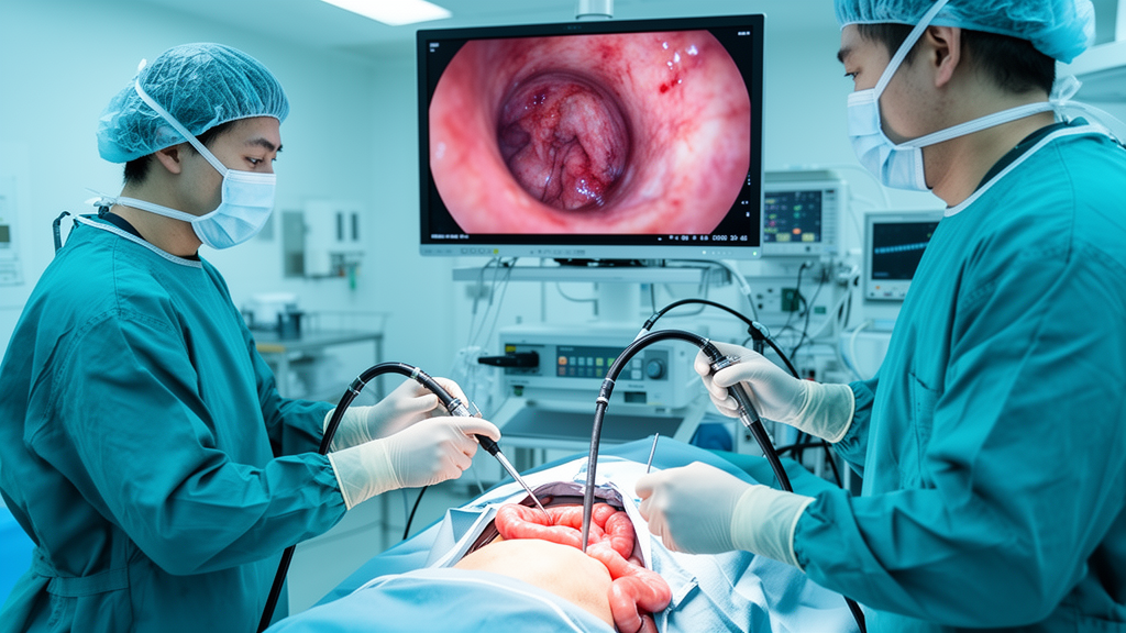
In a newly reported clinical investigation, clinicians utilized intraoperative enteroscopy during exploratory laparoscopy to directly inspect the entire small-bowel mucosa and guide tailored segmental surgical resection in a 58-year-old Han Chinese man suffering from cryptogenic multifocal ulcerous stenosing enteritis. Cryptogenic multifocal ulcerous stenosing enteritis, commonly abbreviated as CMUSE, represents an exceptionally rare small intestinal disorder of unclear etiology that is characterized by multiple superficial ulcerations and short-segment circumferential fibrotic strictures. Because the pathologic lesions in CMUSE remain predominantly discontinuous and strictly confined to the mucosal and submucosal layers of the intestinal wall, conventional non-invasive cross-sectional radiologic imaging and standard endoscopic modalities frequently fail to delineate the full longitudinal extent and exact distribution of active disease. This single-patient case study highlights how introducing a conventional endoscope through a limited surgical enterotomy allowed operating surgeons to visualize four distinct strictured segments across the entire small intestine from the ligament of Treitz to the ileocecal valve, resolving diagnostic uncertainties that had persisted throughout two decades of recurrent bowel obstructions.
The patient presented with a twenty-year clinical history of recurrent abdominal pain and intermittent small-bowel obstruction episodes that had repeatedly required emergency hospitalizations and conservative stabilization. Typical obstructive episodes were marked by severe colicky pain localized to the right lower quadrant, characteristically developing at night and accompanied by prominent abdominal distension as well as the complete cessation of flatus and defecation. Severe vomiting accompanied these recurrent episodes, presenting initially with gastric contents and progressing to bile-stained fluid during particularly protracted attacks. Each prior episode had gradually resolved over several days with conservative inpatient care, including bowel rest, fluid replacement, and gastric acid suppression, but no definitive etiologic diagnosis had ever been achieved across twenty years of medical evaluation. During the month leading up to his definitive admission, the frequency and severity of the painful episodes escalated substantially, accompanied by persistent postprandial vomiting and an unintentional body weight loss of approximately five kilograms, prompting referral for specialized diagnostic workup and surgical evaluation.
A thorough clinical review established the patient had a forty-year history of cigarette smoking and chronic hepatitis B virus infection with positive hepatitis B surface antigen and documented low-level viremia, while testing negative for hepatitis C virus and human immunodeficiency virus. He reported no history of alcohol consumption and denied any chronic or intermittent usage of aspirin, non-steroidal anti-inflammatory drugs, or over-the-counter analgesics. Comprehensive questioning revealed no history of arthritis, chronic musculoskeletal disorders, or inflammatory joint pain that would necessitate analgesic therapy. His sole remote analgesic exposure consisted of a brief one-to-two-week course of an over-the-counter analgesic taken two years prior to admission following surgical fixation of a traumatic left shoulder fracture, with original prescription records no longer retrievable. The patient had undergone no previous abdominal surgeries, and there was no personal or family history of inflammatory bowel disease, systemic autoimmune disorders, or gastrointestinal malignancies, reinforcing the diagnostic complexity of his long-standing obstructive intestinal symptoms.
Upon physical examination at hospital admission, the patient remained hemodynamically stable with a body temperature of 36.7 degrees Celsius, a heart rate of 58 beats per minute, a respiratory rate of 18 breaths per minute, and a blood pressure of 110/72 mmHg, presenting with a body weight of 44.85 kilograms and a height of 158 centimeters. The physical examination of the abdomen was non-tender and soft without palpable masses, guarding, or abnormal auscultatory bowel sounds. Routine laboratory tests demonstrated mild hypoalbuminemia with a serum albumin level of 39.2 grams per liter and a positive fecal occult blood test, whereas inflammatory and autoimmune markers were unremarkable. The patient’s erythrocyte sedimentation rate was normal at 6 millimeters per hour, routine serum tumor markers were within normal laboratory reference ranges, and a comprehensive systemic autoimmune screening panel including antinuclear antibodies yielded negative results. Initial management consisted of complete bowel rest, intravenous fluid resuscitation, and antispasmodic analgesia, which stabilized the patient within two days without requiring nasogastric decompression.
The medical team initiated extensive diagnostic evaluations to localize the source of recurrent obstruction, beginning with contrast-enhanced computed tomography enterography of the small intestine. The CT enterography scan demonstrated localized wall thickening and abnormal mural enhancement within the ileum, suggesting active localized inflammation with luminal narrowing. To visually assess the small intestine, physicians carried out conventional esophagogastroduodenoscopy and colonoscopy, both of which showed completely normal mucosal architecture without identifiable ulceration. Subsequently, bidirectional double-balloon enteroscopy was attempted using both anterograde oral and retrograde anal approaches. However, the specialized enteroscopy scopes were unable to reach the diseased ileal segment or achieve total enteroscopy, leaving the suspected strictures unreachable and uncharacterized by conventional intraluminal visualization. Magnetic resonance enterography was considered as a radiation-free alternative capable of dynamic functional assessment, but because computed tomography had already confirmed the ileal abnormality, mucosal visualization remained the overriding unmet clinical priority.
Because comprehensive non-operative endoscopic and radiologic examinations failed to define the lesions, capsule endoscopy was considered after multidisciplinary consultation and extensive counseling regarding the recognized hazards of device retention. Patency capsule testing was not available at the treating medical institution at that time, precluding pre-procedure mechanical confirmation of intestinal luminal patency. Although guideline protocols caution against deploying video capsules in patients with suspected intestinal stricturing without established luminal patency, the examination was undertaken with the understanding that capsule retention would localize the obstruction site. During the examination, the ingested capsule successfully transmitted images of two distinct stenosing ulcerations before becoming impacted between them, failing to pass spontaneously through the ileal lumen. While the retained video capsule confirmed the presence of obstructive mucosal strictures, it remained trapped, leaving the precise total count, longitudinal distribution, and proximal or distal boundaries of additional strictures unknown prior to surgical intervention.
Given the persistent obstructive pathology, preoperative diagnostic suspicion of CMUSE, and acute capsule impaction, the surgical team proceeded with exploratory laparoscopy. Laparoscopic visualization revealed marked segmental wall thickening accompanied by dense perilesional adhesions situated approximately 80 centimeters proximal to the ileocecal valve, alongside marked dilation of the upstream small bowel. Following laparoscopic adhesiolysis, the surgeons made an 8-centimeter lower midline incision to exteriorize the affected ileal loops. A controlled 2-centimeter enterotomy was performed directly at the site of capsule impaction, allowing the retrieval of the retained device. A standard gastroscope was then introduced through this enterotomy to perform comprehensive intraoperative enteroscopy, thoroughly inspecting the entire mucosal surface of the small intestine from the ligament of Treitz down to the ileocecal valve. This direct mucosal survey revealed four separate circumferential strictures with superficial ulcerations separated by macroscopically normal intervening mucosa, identifying two additional active strictures that had escaped preoperative detection.
Based on the real-time intraluminal mapping provided by intraoperative enteroscopy, the surgical team established clear resection margins ten centimeters proximal and ten centimeters distal to the diseased bowel segment. The entire clustered pathological area encompassed approximately forty centimeters of small intestine, allowing the surgeons to perform a single, tailored en-bloc segmental resection measuring approximately sixty centimeters in total length. This targeted strategy ensured that all four circumferential strictures and associated ulcerations were completely excised while preserving uninvolved small intestine. Intestinal continuity was promptly reconstructed by performing a side-to-side stapled anastomosis with a linear cutting stapler, reinforced with interrupted 3-0 absorbable sutures, after which the mesenteric defect was carefully closed. The remaining jejunum and ileum demonstrated no evidence of obstruction or luminal compromise, and the bowel was arranged and lightly anchored with interrupted absorbable sutures to prevent acute postoperative angulation or mechanical kinking.
Histopathological evaluation of the resected intestinal specimen confirmed the presence of multiple superficial ulcerations strictly confined to the mucosal and submucosal layers, accompanied by dense submucosal fibrotic stricture formation. Importantly, microscopic examination showed complete absence of transmural inflammatory infiltration, fissuring ulcers, or granuloma formation, effectively ruling out Crohn’s disease. Additional diagnostic exclusions were systematically documented: negative interferon-gamma release assays and absence of caseating granulomas excluded intestinal tuberculosis, negative autoimmune serology and lack of vasculitic changes excluded Behcet’s disease, and histology confirmed no malignant lymphoma or viral inclusion bodies. The pathologists specifically differentiated the tissue changes from non-steroidal anti-inflammatory drug-induced diaphragm disease, noting that although both disorders present with superficial ulceration and muscularis mucosae hyperplasia, the specimen lacked thin web-like fibrous septa, mucosal bridges, or fold-crest ulceration. Furthermore, myenteric plexus hyperplasia was identified, supporting the diagnosis of CMUSE. Although genetic testing for SLCO2A1 mutations associated with chronic enteropathy was recommended, the patient declined testing.
The patient achieved an uneventful postoperative recovery without surgical site infection, anastomotic leakage, or bowel dysmotility, and was discharged in stable clinical condition. At his one-month outpatient follow-up, the patient reported complete freedom from abdominal pain and noted a dramatic restoration of digestive comfort. By the six-month evaluation, his nutritional status had markedly improved, accompanied by a documented five-kilogram weight gain and no recurrent obstructive symptoms. A telephone follow-up at ten months confirmed he remained completely symptom-free. The study authors emphasized that while intraoperative enteroscopy successfully guided resection and uncovered occult strictures in this patient, these single-case observations cannot establish whether the procedure provides a statistically definitive bowel-preserving advantage over conventional surgical inspection alone. Furthermore, the ten-month surveillance period remains brief given recognized long-term CMUSE recurrence risks, and clinicians reiterated that capsule endoscopy without prior patency confirmation poses significant retention risks and should not be used as an intentional localization strategy.
Subject of Research: Intraoperative enteroscopy for cryptogenic multifocal ulcerous stenosing enteritis diagnosis and surgical management
Article Title: Cryptogenic multifocal ulcerous stenosing enteritis diagnosed and managed with intraoperative enteroscopy: a case report
Article References: Yu, P., Zheng, J., Xu, B., & Huang, Y. (2026). Cryptogenic multifocal ulcerous stenosing enteritis diagnosed and managed with intraoperative enteroscopy: a case report. Frontiers in Medicine, 13, Article 1999987. https://doi.org/10.3389/fmed.2026.1999987
Image Credits: AI Generated
DOI: 10.3389/fmed.2026.1999987
Keywords: cryptogenic multifocal ulcerous stenosing enteritis, CMUSE, intraoperative enteroscopy, small bowel obstruction, capsule endoscopy, capsule retention, segmental bowel resection, laparoscopic surgery, submucosal fibrosis, mucosal ulceration, CT enterography, double-balloon enteroscopy, Crohn's disease differentiation, NSAID enteropathy, diaphragm disease, SLCO2A1 gene, enterotomy
News Source: Ophelia Keating. (November 17, 2026). Intraoperative Enteroscopy Successfully Identifies Hidden Strictures in Cryptogenic Enteritis Case. Scienmag.