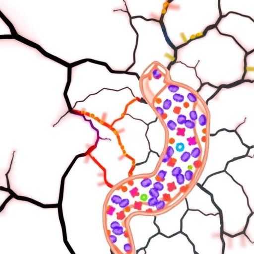
In a groundbreaking study published in Nature, researchers have unveiled the critical role of intestinal macrophages in modulating the progression of synucleinopathy along the gut–brain axis, providing fresh insights into the early stages of Parkinson’s disease (PD). This pioneering work places self-maintaining macrophages in the mesenteric environment (ME-Macs) at the heart of α-synuclein (αS) pathology initiation and propagation, challenging conventional perspectives that predominantly centered on neuronal involvement.
The investigation leverages sophisticated transgenic mouse models expressing pathological αS variants and direct injections of PD-derived αS fibrils into the mesentery to mimic gut-originating αS pathology. Intriguingly, these models reveal that αS aggregation begins within the enteric nervous system (ENS), concomitant with gastrointestinal dysfunction characterized by increased gut transit time. This aligns closely with Braak staging hypotheses, which propose that in body-first PD phenotypes, αS pathology originates in peripheral tissues, such as the gut, before ascending to the central nervous system (CNS).
Key to the process are the ME-Macs, which demonstrate elevated uptake of phosphorylated αS at serine 129 (s129p+), a pathological hallmark. Despite their established role in maintaining ENS homeostasis, these macrophages are shown to harbor significant amounts of misfolded αS, implicating them as both facilitators and modulators of αS protein aggregation. Notably, ME-Macs upregulated autophagic and lysosomal clearance pathways, yet paradoxically appeared to foster a microenvironment conducive to αS aggregate accumulation—highlighting a dualistic function that may underpin early-stage synucleinopathy.
Targeted depletion experiments further illustrate the indispensability of ME-Macs in disease progression. When selectively ablated, these macrophages substantially mitigate the spread of s129p+ αS inclusions from the ENS into the CNS as well as attenuate motor dysfunction in murine models. Despite minor collateral reductions in eosinophil and monocyte populations, the findings underscore ME-Macs as crucial nodes for αS propagation, possibly through their expression of PD-associated risk genes such as Gba1, Lrrk2, Vps35, and Ctsd—genes heavily implicated in lysosomal functioning and protein degradation.
Beyond macrophage-centric mechanisms, the study reveals a potent immunological axis involving T cell expansion. ME-Macs were found to significantly influence the local proliferation of CD3+ T cells in the ENS, which subsequently migrate to CNS regions pertinent to PD neuropathology, notably the dorsal striatum and lateral ventricles. This T cell trafficking phenomenon supports a paradigm where αS pathology incites neuroimmune crosstalk, potentially exacerbating neurodegeneration through sustained inflammatory milieu and antigen presentation dynamics.
Pharmacological intervention with fingolimod, a T cell trafficking inhibitor, demonstrated neuroprotective effects by reducing αS spread and corresponding neurodegeneration in PD mouse models. This not only affirms the pathological role of T cells in synucleinopathy progression but opens avenues for immunomodulatory therapeutic strategies. Nonetheless, the complex, systemic immunoregulatory effects of such drugs warrant further elucidation to decipher specificity within PD pathology.
Elucidating the cellular communication channels, the study highlights that ME-Macs upregulate major histocompatibility complex class II (MHCII) molecules in response to αS pathology, positioning them as probable antigen-presenting entities capable of modulating T cell responses. However, the potential interplay with dendritic cells—known professional antigen presenters—remains an open question. It is conceivable that ME-Macs transfer αS antigens to dendritic cells to facilitate T cell priming, but definitive mechanisms await discovery.
Furthermore, TGFβ signaling within ME-Macs emerged as a pivotal factor in governing T cell expansion triggered by αS. The attenuation of TGFβ expression in ME-Macs corresponds with reduced T cell proliferation, underscoring a cytokine-mediated axis crucial for immune modulation. Given TGFβ’s known influence on T cell differentiation into regulatory or inflammatory phenotypes, its role in synucleinopathy-associated immune dysregulation represents a promising therapeutic target.
The identification of ME-Macs’ unique perivascular gene signatures resembling CNS border-associated macrophages also hints at conserved immune regulatory functions across the gut–brain interface. This resemblance could explain their specialized roles in recruiting and activating CD4+ T cells within neuroimmune niches, a process previously implicated in αS-overexpressing models. It further heightens the need to explore whether such tissue-resident macrophages universally contribute to neurodegeneration via immune mechanisms.
Importantly, this work contextualizes environmental and intrinsic triggers for αS aggregation in human gut tissue, including pesticide exposure and viral infections known to elevate enteric αS expression and phosphorylation. Ageing similarly contributes to progressive αS modifications, suggesting a multifactorial etiology that primes ME-Macs and associated immune cells for pathological engagement. These exogenous and endogenous factors may serve as initial catalysts for the cascade leading to PD.
While the model centers on direct pathogenic αS administration, the study acknowledges the elusive endogenous triggers driving initial αS misfolding events in humans. Future research must decode this initial seeding, illuminating preventive strategies for PD. Integration of known PD risk genes within macrophage lysosomal pathways accentuates the biological convergence toward impaired αS clearance as a fundamental disease mechanism.
Finally, the clinical implications of these findings are profound. By placing ME-Macs at the forefront of αS pathology transmission and immune modulation, the study not only reshapes the understanding of gut–brain axis involvement in PD but also proposes novel diagnostic and therapeutic opportunities. Targeting ME-Macs and their crosstalk with T cells could permit interception of disease progression at early, potentially reversible stages, transforming the landscape of PD management and offering hope for millions globally.
Subject of Research:
Intestinal macrophages and their role in modulating α-synuclein pathology and immune responses along the gut–brain axis in Parkinson’s disease models.
Article Title:
Intestinal macrophages modulate synucleinopathy along the gut–brain axis.
Article References:
De Schepper, S., Konstantellos, V., Conway, J.A. et al. Intestinal macrophages modulate synucleinopathy along the gut–brain axis. Nature (2026). https://doi.org/10.1038/s41586-025-09984-y
Image Credits: AI Generated
DOI: https://doi.org/10.1038/s41586-025-09984-y
Tags: Braak staging hypothesisenteric nervous system dysfunctiongastrointestinal dysfunctiongut-brain axisintestinal macrophagesmesenteric environment macrophagesneuroinflammation and gut healthParkinson’s disease pathologyprotein aggregation in macrophagessynucleinopathy progressiontransgenic mouse modelsα-synuclein aggregation

