
In the realm of nanoscale analysis, cryo-transmission electron microscopy (cryo-TEM) has long been a critical tool for observing biological and soft materials in conditions that preserve their innate structures. By rapidly freezing samples within a solvent, cryo-TEM enables scientists to capture images that closely mirror the native state of delicate specimens, circumventing the distortions or damage typically induced by traditional preparation methods. This technique provides crucial insights into the morphology, size, and dispersion of nanoparticles and biological macromolecules embedded in their aqueous environments. Despite its impressive capabilities, until recently, cryo-TEM has struggled to reveal the elemental composition of these organic nanomaterials accurately, leaving a significant gap in comprehensive structural analysis.
The elemental makeup of materials plays a decisive role in understanding their chemical behaviors, functions, and interactions. Standard approaches such as energy-filtered transmission electron microscopy (EF-TEM), coupled with electron energy loss spectroscopy (EELS), have offered pathways to elemental mapping. However, the application of these methods to soft, organic substances has been hampered by several formidable challenges. Chief among these are beam-induced sample damage and image blurring induced by sample drift during the prolonged acquisition times required for EELS mapping. The inherent fragility of organic nanomaterials and biological specimens makes them particularly susceptible to these limitations. Moreover, conventional EF-TEM techniques have predominantly been confined to inorganic or metallic samples or large-area specimens, thereby restricting their utility in biomedical and organic material research.
Addressing these critical barriers, a team of researchers at Tohoku University has pioneered an innovative elemental mapping technique that marries cryo-TEM with EELS and EF-TEM under low-dose conditions. This breakthrough enables simultaneous, high-resolution visualization of both the structural attributes and elemental distributions of nanomaterials submerged in frozen solvents. Their approach leverages a sophisticated integration of techniques to preserve sample integrity, minimize drift, and enhance signal clarity, opening new vistas for detailed analyses of organic and bio-related materials at the nanoscale with unprecedented accuracy.
.adsslot_kNhVc3MK2b{width:728px !important;height:90px !important;}
@media(max-width:1199px){ .adsslot_kNhVc3MK2b{width:468px !important;height:60px !important;}
}
@media(max-width:767px){ .adsslot_kNhVc3MK2b{width:320px !important;height:50px !important;}
}
ADVERTISEMENT
One of the pivotal obstacles the team sought to overcome was the interference caused by plasmon excitations originating from the vitreous ice matrix encapsulating the samples. These plasmon signals heightened the spectral background noise substantially, thereby obscuring the true elemental signatures of the organic nanoparticles under scrutiny. Recognizing this confounding factor, the researchers implemented an advanced imaging strategy that couples the established 3-window method—a technique for precise background subtraction in EELS—with state-of-the-art drift compensation algorithms. This dual installation effectively isolates the sample’s elemental signals from the overwhelming background, resulting in crisp, clear elemental maps that faithfully represent the native material composition.
Integral to this innovation was the development of bespoke software enhancements for the “ParallEM” electron microscope control system. The new program automates and refines control over energy shifts during data capture, facilitating a streamlined imaging workflow that ensures consistent data integrity and repeatability. By managing energy compensations dynamically, the system effectively counteracts the subtle energetic variations during prolonged scans, which would otherwise blur or distort elemental maps. This technical advancement not only enhances image fidelity but also promotes user-friendly operation, encouraging broader adoption in materials research laboratories.
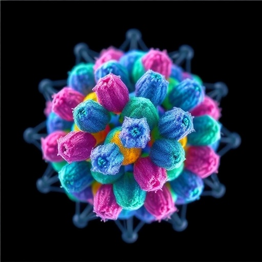
The practical impact of this technique was demonstrated through elemental mapping of silica nanoparticles embedded in frozen solvents, a challenging test case due to the light atomic weight elements involved. Using cryo-EELS/EF-TEM, the researchers successfully mapped silicon distributions with remarkable spatial resolution, vividly delineating the structure and dispersion profiles of individual silica nanoparticles. Their methodology achieved a detection threshold at particle sizes as small as approximately 10 nanometers, marking a significant improvement in sensitivity compared to traditional elemental mapping approaches.
Expanding the scope of their method’s applicability, the team investigated hydroxyapatite nanoparticles, which constitute the primary inorganic mineral component of human bones and teeth. The elemental maps revealed precise spatial distributions of calcium and phosphorus within these particles, two biologically significant elements that are fundamental to understanding bone mineralization and related physiological processes. This application underscores the method’s potential to unravel complex compositional heterogeneities in biominerals, potentially advancing fields such as biomaterials engineering and medical diagnostics.
The implications of this work resonate far beyond the immediate cases studied. By significantly reducing electron doses and mitigating sample drift while achieving high spatial resolution elemental mapping, this new cryo-EELS/EF-TEM approach stands poised to revolutionize the analysis of a broad spectrum of materials. From biomolecules and medical implants to catalysts and industrial inks, the ability to probe both structure and elemental composition concurrently and non-destructively opens exciting avenues for innovation in life sciences, chemistry, materials science, and nanotechnology.
Future research leveraging this low-dose elemental mapping promises to deepen insights into nanoscale phenomena that govern material functionalities and interactions. For instance, in catalysis, understanding the precise distribution of active elements within nanoparticles could optimize efficiency and selectivity. In the biomedical arena, mapping trace elements in cells or tissues can illuminate pathological processes or inform novel therapeutic strategies. The combination of cryogenic preservation and sensitive elemental analysis thus ushers in a new era of correlative microscopy that integrates chemical and structural data at the nanoscale.
Beyond the technical achievements, the Tohoku University team’s approach also highlights the importance of interdisciplinary collaboration and engineering ingenuity. By integrating physics, materials science, software development, and microscopy expertise, they have crafted a platform that transcends the traditional limits of electron microscopy applied to soft matter. The new technique’s robustness and adaptability ensure it can be tailored to various experimental setups and sample types, thereby enhancing its potential impact across research communities worldwide.
With the details of their research published in Analytical Chemistry, this advancement has already begun to permeate the scientific community, inviting further exploration and refinement. The publication details the comprehensive methodology, experimental validations, and technical considerations pivotal to reproducing and extending this technique. It represents a significant step toward routine elemental imaging of organic nanomaterials in their native hydrated states, bridging a longstanding gap in microscopy and spectroscopy.
As we continue to explore the nanoscale world, techniques such as low-dose cryo-EELS/EF-TEM will be indispensable for elucidating the subtle chemical complexities embedded within fragile biological and soft materials. The capacity to capture these intricate details without inflicting structural or compositional damage positions this technology at the forefront of modern materials characterization. Its potential to drive discoveries and innovations across multiple disciplines underscores the ever-evolving synergy between imaging technology and scientific inquiry.
Subject of Research: Novel low-dose elemental mapping technique for organic and bio-related nanomaterials using cryo-EELS/EF-TEM.
Article Title: Low-Dose Elemental Mapping of Light Atoms in Liquid-phase Materials Using Cryo-EELS
News Publication Date: 31-Jul-2025
Web References:
http://dx.doi.org/10.1021/acs.analchem.5c02121
Image Credits: ©Daisuke Unabara et al.
Keywords
Cryo electron microscopy, Microscopy, Nanomaterials, Organic matter, Chemistry, Materials science
Tags: advances in nanoscale imagingbeam-induced damage in electron microscopychallenges in cryo-TEM analysiscryo-transmission electron microscopy applicationselectron energy loss spectroscopy methodselemental distribution in biological specimenselemental mapping in nanomaterialsimaging techniques for soft materialsinnovative imaging techniques for materials sciencenanoparticle morphology analysisorganic nanomaterials characterizationpreserving native structures in microscopy


