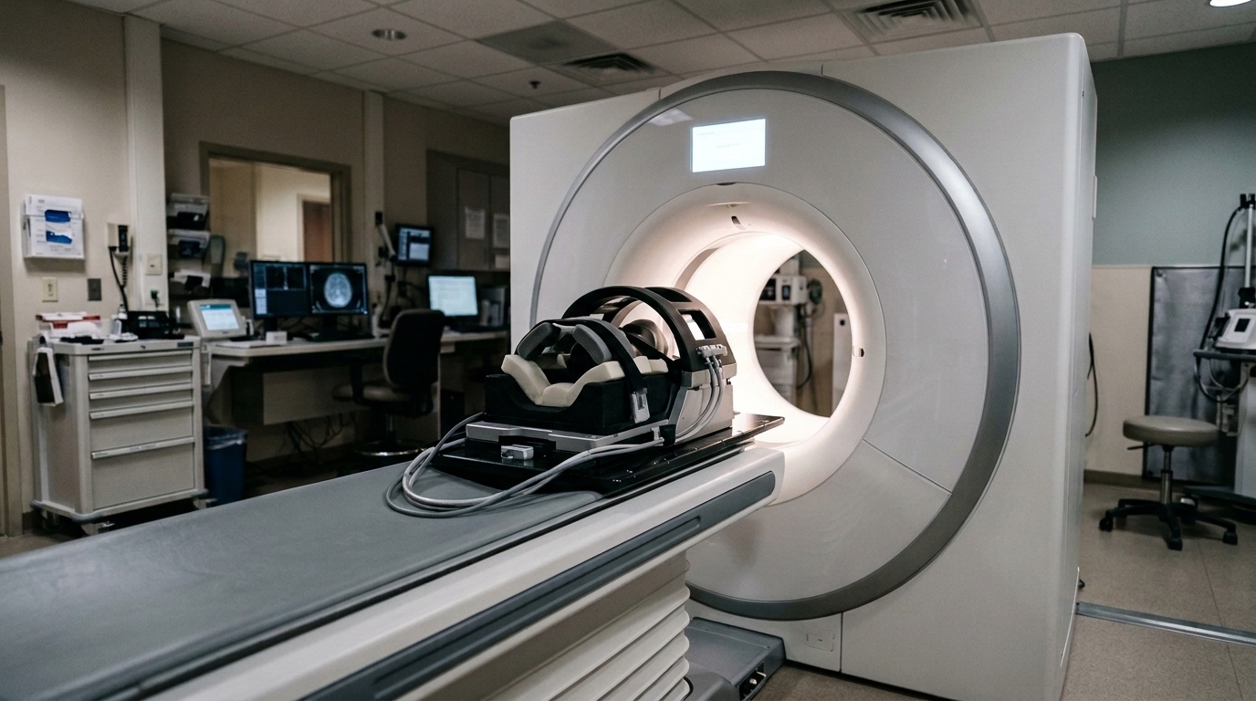
An improved 2.5D-UNet deep learning architecture enhances the accuracy of pseudo-computed tomography images generated from non-attenuation-corrected positron emission tomography scans, according to an original research study published in EJNMMI Physics. By utilizing adjacent image slices to provide contextual anatomical information, the model enables attenuation correction without requiring additional radiation exposure or dedicated imaging equipment.
In positron emission tomography, tissue attenuation represents a significant imaging challenge that can lead to quantification errors, the underestimation of lesions, and the misinterpretation of the extent of disease. Correcting for attenuation is commonly achieved using a computed tomography scan, which provides the necessary anatomical details. However, relying on computed tomography substantially increases the radiation dose received by patients, raises imaging equipment expenses, and can introduce misregistration errors between the scans.
To overcome these clinical limitations, deep learning methods have been introduced to synthesize pseudo-computed tomography images directly from non-attenuation-corrected scans. In this work, researchers developed an improved 2.5D-UNet architecture designed to process three consecutive slices to provide enriched contextual information for generating a single slice. The model was trained using a dataset consisting of 189 non-attenuation-corrected head scans from patients injected with either 18F-fluorodeoxyglucose or 11C-acetoacetate radiotracers, paired with corresponding computed tomography images.
Evaluating the generated pseudo-computed tomography images across all patients revealed overall performance gains with the proposed 2.5D network over a standard 2D architecture. The soft tissue Dice score improved from 0.905 ± 0.015 with the 2D model to 0.922 ± 0.017 with the 2.5D approach. In addition, the structural similarity index measure increased from 0.889 ± 0.023 in the 2D configuration to 0.921 ± 0.019 using the 2.5D framework.
Beyond slice-level numerical metrics, three-dimensional forward projections comparing total attenuation projection values between actual computed tomography scans and synthesized pseudo-computed tomography images highlighted the superior attenuation maps produced by the 2.5D deep learning framework. The authors concluded that incorporating adjacent slices for contextual guidance allows the 2.5D-UNet to outperform 2D models, delivering fast and effective attenuation correction from non-attenuation-corrected head scans.
Subject of Research: Deep learning-based pseudo-CT generation for PET attenuation correction
Article Title: Improved 2.5D-UNet pseudo-CT generation for attenuation correction in PET imaging using 18F-fluorodeoxyglucose and 11C-acetoacetate radiotracers
Article References: St-Georges, A., Houle, A., Cenci, T., Giroux, J.-S., Toussaint, M., Thibaudeau, C., Auger, E., Croteau, É., Cunnane, S., Lecomte, R., & Michaud, J.-B. (2026). Improved 2.5D-UNet pseudo-CT generation for attenuation correction in PET imaging using 18F-fluorodeoxyglucose and 11C-acetoacetate radiotracers. EJNMMI Physics. https://doi.org/10.1186/s40658-026-00909-w
Image Credits: AI Generated
DOI: 10.1186/s40658-026-00909-w
Keywords: PET imaging, attenuation correction, pseudo-CT, deep learning, 2.5D-UNet, radiotracers, medical imaging, head scans, Improved, D-UNet, generation, attenuation
News Source: Ophelia Keating. (November 7, 2026). Improved 2.5D-UNet Enhances Pseudo-CT Generation for Head PET Attenuation Correction. Scienmag.