For over a century, vasomotionâthe rhythmic contraction and relaxation of blood vessel wallsâhas intrigued scientists for its subtle yet critical role in regulating blood flow. This dynamic oscillation, particularly important in the brain’s intricate vascular network, is thought to underpin essential physiological processes ranging from fine-tuning cerebral perfusion to waste clearance. Despite its known associations with neurological health and diseases such as Alzheimerâs and stroke, the precise mechanisms and spatiotemporal characteristics of vasomotion within the living brain have remained elusive. Conventional imaging techniques lacked the temporal resolution or the spatial breadth to map these fleeting vascular rhythms with precision.
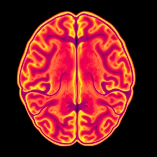
Recently, a breakthrough study from Aarhus University has shed transformative light on the origin and propagation of vasomotion in the awake mouse brain. Published in the journal Neurophotonics, this research employed laser speckle contrast imaging (LSCI), a cutting-edge optical technique that captures real-time, high-resolution maps of blood flow across extensive cortical regions. LSCI leverages the interference patterns, or âspeckles,â formed by coherent laser light scattered by moving red blood cells, providing a non-invasive window into cerebral hemodynamics at both microscopic scales and wide fields of view. By harnessing this technology in awake, non-anesthetized mice, researchers sidestepped the confounding effects of anesthesia, preserving physiological vascular dynamics.
Central to their approach was the integration of sophisticated computational analyses. Wavelet transforms allowed the team to dissect blood flow signals into time-frequency components, pinpointing the characteristic vasomotion frequency bands with unprecedented accuracy. Meanwhile, a customized “pulsatility index” algorithm enabled the discrimination between arterial and venous vessels based on their distinctive flow pulsations. Advanced clustering algorithms then parsed the continuous data streams to isolate bursts or “flares” of vasomotion activity, distinguishing periods of vascular oscillations from silent intervals.
Their findings revealed that vasomotion is inherently transient rather than continuous, manifesting as discrete episodes lasting roughly 80 seconds, punctuated by silent gaps of similar duration. Intriguingly, these oscillatory bursts originate predominantly from the walls of small arteries. Here, vessel diameter exhibited rhythmic fluctuations, which then propagated downstream as pulsatile waves altering blood flow. Tracking these dynamic signals unveiled a remarkable delay of approximately 0.3 seconds before the oscillations appeared in draining veins, demonstrating a clear directional flow of vasomotor signals through the vascular tree.
This spatially and temporally resolved portrait elucidates a fundamental physiological mechanism: arterial walls actively generate vasomotion pulses that ripple through the cerebrovascular network, modulating flow in a coordinated, wave-like manner. This challenges prior assumptions that vasomotion might arise diffusely or simultaneously across vessels and highlights arterial smooth muscle cellsâ critical role in orchestrating these rhythms. The transient and intermittent nature of vasomotion underscores its possible function as a responsive, adaptive regulator rather than a constant driver of cerebral blood flow.
The implications of this work extend beyond basic vascular physiology. Given vasomotionâs putative role in facilitating the clearance of metabolic waste via perivascular pathways, understanding its dynamics offers potential new insights into how impaired blood flow patterns may contribute to neurodegenerative diseases. Disruptions in the timing or propagation of vasomotor waves might underlie pathological states where blood perfusion and brain homeostasis falter. By mapping the precise temporal bursts and spatial origins of vasomotion, future therapeutic approaches could aim to restore or modulate these essential vascular rhythms.
Commenting on the study, Alberto L. Vasquez from the University of Pittsburghâs Center for Neuroscience emphasized the cleverness of using LSCI for this application. He noted that this method reveals not only the amplitude but also frequency variations of slow vascular pulsations across the awake brain in high detail, suggesting potential control points along vessels. This high-resolution perspective opens avenues for identifying focal sites of vascular regulation and the downstream effects of localized oscillations.
This study exemplifies the power of combining state-of-the-art optical imaging with advanced signal processing to decode complex neurovascular phenomena. By capturing the brain’s dynamic vascular landscape in action, the researchers have opened a new window into cerebrovascular function that balances wide-field observation with temporal precision. The ability to chronicle the ebb and flow of vascular oscillations in awake, behaving animals bridges a crucial gap in our understanding of how blood supply is finely tuned in real physiological states.
Moreover, the approach described paves the way for future investigations into how systemic factors, neural activity, or pathological conditions influence vasomotion patterns. It invites exploration of how neurovascular couplingâthe relationship between neuronal demands and vascular responsesâis modulated through these rhythmic vessel contractions. Integrating these vascular signals with concurrent neuronal recordings may unravel deeper layers of brain function and its vascular underpinnings.
The work also illustrates the critical role of arterial smooth muscle dynamics in shaping cerebral blood flow, adding to a growing body of evidence that vascular tone is not merely a passive backdrop but an active player in brain health. It challenges researchers to consider transient, wave-like phenomena in vascular biology rather than static or uniform states. Such a paradigm shift could have profound implications in diagnosing, monitoring, and treating cerebrovascular disorders.
In addition to providing fundamental scientific insights, this research highlights the translational potential of LSCI and computational analytics for clinical applications. Non-invasive imaging techniques with the temporal and spatial fidelity demonstrated here could one day inform patient-specific assessments of vascular function or detect early signatures of vascular dysregulation in neurodegeneration.
Overall, the study marks a milestone in vascular neuroscience, illuminating how pulsatile activity arises and traverses the cerebral vascular network. By revealing the arterial wall origin and transient nature of vasomotion, it deepens our grasp of cerebral blood flow regulation, setting the stage for innovative research into vascular health and disease.
Subject of Research: Cerebrovascular dynamics, vasomotion, and flow regulation in the awake mouse brain.
Article Title: Arterial-wall origin and transient dynamics of flow- and vaso-motion activities in the awake mouse brain revealed by laser speckle contrast imaging
News Publication Date: 19-Aug-2025
Web References:
Original article in Neurophotonics
SPIE Journal link DOI: 10.1117/1.NPh.12.S2.S22804
References:
M. V. Skøtt, V. Matchkov, and D. D. Postnov, âArterial-wall origin and transient dynamics of flow- and vaso-motion activities in the awake mouse brain revealed by laser speckle contrast imaging,â Neurophotonics 12(S2), S22804 (2025).
Image Credits:
M. V. Skøtt et al., doi 10.1117/1.NPh.12.S2.S22804.
Keywords:
High resolution imaging, In vivo imaging, Neuroimaging, Vascular biology, Brain
Tags: advances in neurophotonics for brain studiesblood vessel dynamics in awake micehigh-resolution imaging of cerebral hemodynamicsimaging techniques for cerebral blood flowimplications of vasomotion for Alzheimer’s and stroke researchlaser speckle contrast imaging in neuroscienceneurological health and blood flow regulationnon-invasive brain imaging methodsphysiological roles of cerebral perfusionreal-time mapping of brain vascular rhythmsunderstanding vasomotion in neurological diseasesvasomotion in brain blood vessels
