In a groundbreaking advancement poised to revolutionize cardiovascular diagnostics, scientists have developed a hypersensitive method capable of detecting single millimeter-sized vascular emboli in vivo with unprecedented precision. Published recently in Nature Communications, this pioneering research delivers a transformative approach to identifying even the smallest and most elusive emboli—adhesive clots that travel through the bloodstream and pose severe health risks such as strokes, heart attacks, and pulmonary embolisms. The potential to detect these dangerous obstructions before they escalate could revolutionize clinical outcomes and save countless lives worldwide.
At the heart of this breakthrough lies an innovative detection technique that leverages enhanced imaging protocols and novel biochemical markers to pinpoint emboli as small as one millimeter—dimensions previously challenging to identify within the complex vascular network of living organisms. The ability to observe these microscopic obstructions in real-time and in a non-invasive manner represents a critical leap forward, as traditional imaging modalities like computed tomography angiography and magnetic resonance imaging often fall short when it comes to detecting diminutive embolic formations. These conventional limitations can delay intervention, resulting in dramatic consequences for patients.
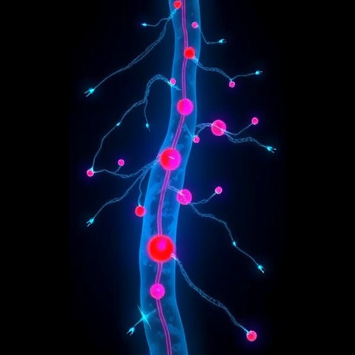
The research team, led by Liu, R., Li, S., and Gao, X., meticulously engineered an integrative platform combining advanced fluorescence molecular imaging with specialized adhesive tracers designed to bind selectively to emboli surfaces under physiological conditions. This biochemical refinement enhances signal specificity dramatically, allowing for unambiguous visualization of emboli in vivo without interference from surrounding vascular structures or circulating blood cells. By optimizing the adhesive properties of these tracers, the researchers enabled them to adhere robustly to emboli, amplifying detection sensitivity to levels not previously achievable.
Central to the method’s efficacy is its harnessing of near-infrared (NIR) fluorescence, which penetrates deeply through biological tissues while minimizing autofluorescence and background noise. The conjugation of adhesive molecules to NIR fluorophores creates a visually striking contrast when emboli are present, facilitating effortless differentiation from normal blood flow and nearby tissues. This advancement not only aids clinicians in spotting emboli promptly but may also pave the way for real-time monitoring during surgical or interventional procedures, significantly enhancing patient safety.
In addition to the imaging modality, the research highlights the importance of adhesive chemistry tailored specifically to vascular dynamics. The unique environment inside blood vessels, characterized by constant shear forces and shear stress, necessitates tracers with rheological properties capable of sustained attachment. The newly synthesized adhesive compounds exhibit remarkable resistance to detachment even under turbulent flow, ensuring reliable emboli labeling throughout diagnostic examinations.
Equally compelling is the platform’s versatility, as it can be adapted to target a variety of embolic materials, including platelet-rich clots, lipid-based obstructions, and fibrin accumulations. This multiplexing ability is a significant stride toward comprehensive emboli detection across different pathological conditions. Moreover, the platform’s minimally invasive approach obviates the need for biopsies or catheter insertions traditionally used to identify embolic events, cutting down risks and discomfort for patients.
The implications of this discovery extend far beyond diagnostic precision. With the capacity to detect emboli at such an early and localized stage, healthcare providers could initiate prophylactic interventions earlier than ever before. For example, anticoagulant therapies might be administered preemptively upon image-confirmed emboli presence, potentially halting progression to full vascular occlusion. Furthermore, this technology may enable personalized assessment of embolic load in chronic cardiovascular diseases, offering tailored therapeutic regimens based on direct visualization metrics.
From a research perspective, the hypersensitive detection method opens new horizons in understanding emboli dynamics and their role in disease pathogenesis. Real-time visualization of emboli formation, migration, and resolution within living organisms affords unprecedented opportunities for studying the intricate interplay between blood components, endothelial surfaces, and hemodynamic forces. Such insights could catalyze the development of next-generation pharmaceuticals explicitly designed to disrupt emboli genesis or promote their clearance.
Notably, the research exemplifies the power of interdisciplinary collaboration, uniting expertise across molecular biology, bioengineering, chemistry, and clinical sciences to surmount longstanding barriers in vascular imaging. The innovative adhesive molecules represent the fruits of synthetic chemistry ingenuity, while the sophisticated imaging protocols demonstrate cutting-edge advances in optical physics. Furthermore, the translational potential underscores the importance of tightly integrating fundamental research with clinical imperatives.
Technological scalability and accessibility also factor prominently in the study’s significance. The platform’s reliance on fluorescence imaging leverages existing clinical imaging infrastructure present in many hospitals, facilitating potential adoption without exorbitant costs or complex retooling. As the tracers and imaging protocols undergo further refinement, widespread clinical deployment could become feasible, democratizing access to early emboli detection worldwide—even in resource-constrained settings.
However, several challenges remain before broad clinical implementation can be realized. Long-term biocompatibility of the adhesive tracers requires ongoing evaluation to rule out unintended immune reactions or toxicity. Additionally, large-scale clinical trials must validate the sensitivity and specificity metrics observed in preclinical models across diverse patient populations. Addressing these hurdles will be critical for translating this promising technology into routine medical practice.
Looking ahead, researchers are optimistic about expanding the platform’s capabilities by integrating artificial intelligence (AI) algorithms capable of automated emboli recognition and quantification from imaging data. Such integration could streamline clinical workflows, reduce diagnostic errors, and provide continuous monitoring through wearable or implantable devices, heralding a new era of precision cardiovascular medicine. Moreover, coupling the detection technique with targeted drug delivery offers prospects for localized emboli dissolution, minimizing systemic side effects.
The broader impact of this innovation extends to public health, particularly in mitigating the burden of cardiovascular diseases—the leading cause of mortality globally. Early emboli detection aligns synergistically with preventive medicine initiatives, potentially reducing hospital admissions, long-term disabilities, and healthcare expenditures associated with embolic events. By shining light on hidden vascular threats within living organisms, this methodology empowers clinicians with a powerful tool to safeguard patients proactively.
In sum, the hypersensitive detection of single millimeter vascular emboli from adhesive in vivo represents a landmark achievement that confronts critical challenges in cardiovascular diagnostics and interventions. By combining sophisticated adhesive chemistry with advanced fluorescence imaging techniques, the researchers have unlocked a new dimension of vascular visualization that promises profound clinical and scientific dividends. As the medical community embraces and refines this technology, transformative improvements in patient outcomes and disease understanding are poised to follow, ushering in a safer and more informed future for cardiovascular care.
Subject of Research: Detection of millimeter-sized vascular emboli using hypersensitive adhesive probes and fluorescence imaging in living organisms.
Article Title: Hypersensitive detection of single millimeter vascular emboli from adhesive in vivo.
Article References:
Liu, R., Li, S., Gao, X. et al. Hypersensitive detection of single millimeter vascular emboli from adhesive in vivo. Nat Commun (2026). https://doi.org/10.1038/s41467-026-68534-w
Image Credits: AI Generated
Tags: biochemical markers for embolicardiovascular diagnostics advancementsclinical outcomes improvementearly detection of strokes and heart attackshypersensitive detection of vascular emboliin vivo imaging techniquesinnovative imaging protocols in medicinemillimeter-sized emboli detectionNature Communications research findingsnon-invasive emboli identificationreal-time imaging of blood clotstransformative health diagnostics


