Researchers at the Centre for Genomic Regulation (CRG) in Barcelona and the Spanish National Cancer Research Centre (CNIO) in Madrid have captured the world’s first high-resolution images of the earliest moments of microtubule formation inside human cells. The findings, published today in the journal Science, lay the foundations for potential breakthroughs in treating many different types of diseases ranging from cancer to neurodevelopmental disorders.
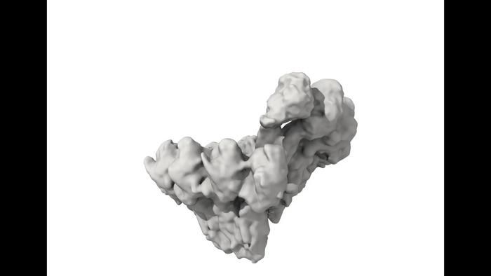
Credit: Marina Serna/CNIO
Researchers at the Centre for Genomic Regulation (CRG) in Barcelona and the Spanish National Cancer Research Centre (CNIO) in Madrid have captured the world’s first high-resolution images of the earliest moments of microtubule formation inside human cells. The findings, published today in the journal Science, lay the foundations for potential breakthroughs in treating many different types of diseases ranging from cancer to neurodevelopmental disorders.
“Microtubules are critical components of cells, but all the images we see in textbooks describing the first moments of their creation are models or cartoons based on structures in yeast. Here we capture the process in action inside human cells. Now that we know what it looks like, we can explore how it’s regulated. Given the fundamental role of microtubules in cell biology, this could eventually lead to new therapeutic approaches for a wide range of disorders,” explains ICREA Research Professor Thomas Surrey, main co-author of the study and researcher at the Centre for Genomic Regulation.
Molecular ‘highways’ of the cell
A cell is much like a bustling city, requiring state-of-the-art infrastructure to function. One of the most important components are microtubules, tubes made of proteins which act like bridges or roads that help move things around and give the cell its shape. Importantly, they are critical for cell division, ensuring that two new cells can be born from a parent cell. In neurons, they are absolutely essential, forming highways for transport over long distances.
Microtubules are built by a large assembly of proteins known as the gamma-tubulin ring complex (γ-TuRC). The proteins work like a blueprint, laying down tiny building blocks called tubulins in a specific order. This is a process called microtubule nucleation, which is like laying the foundation stones of a bridge. Once the foundation is set, tubulins are added to make the bridge as long as necessary.
For the cell to work correctly, microtubules need to be made of thirteen different rows of tubulins. A few years ago, researchers were baffled to discover that human γ-TuRC exposes fourteen rows of tubulins. This was confusing because researchers expected it to be a perfect template for microtubules, which did not seem to be the case. But high-resolution structures had only been pictured of either γ-TuRC or microtubules in isolation and never together – until now.
“We had to find conditions that allowed us to image over a million microtubules in the process of nucleation before they grow too long and obscure the action of γ-TuRC. We were able to achieve this using the molecular toolbox of our lab and then freeze the microtubule stubs in place”, explains Cláudia Brito, postdoctoral researcher at the CRG and co-first author of the study.
High-resolution imaging
To observe γ-TuRC while it was actively forming microtubules, researchers prepared samples at the CRG in Barcelona and the Electronic Microscopy Center at ALBA (EMCA), where they were flash-frozen in a thin layer of ice – preserving the natural shape of the molecules involved and helping discern fine details of structures at near atomic level. Frozen samples were then sent to the Basque Resource for Electron Microscopy (BREM) in Vizcaya, where the high-resolution data generated was then transferred to be analysed at the CNIO in Madrid. Marina Serna, Staff Scientist at CNIO and co-first author of the study, used the images obtained by cryo-electron microscopy and complex image processing methods to determine the 3D structure of γ-TuRC while forming microtubules.
This analysis revealed that as γ-TuRC starts the nucleation process and as the microtubule begins to form, it cleverly changes its shape. Initially in an open state, it progressively closes as the microtubule grows. The change makes γ-TuRC stow away one of its 14 tubulins, effectively matching the design of the microtubule that needs only 13 rows. The whole process is facilitated by a newly-discovered latch mechanism, revealing that it’s the growing microtubule itself which helps the template find its correct shape.
Oscar Llorca, Director of the Structural Biology program at CNIO and main co-author of the paper, explains: “We have visualized the process that initiates microtubule formation, and we see that human γ-TuRC is an open ring that closes to effectively become a perfect template to nucleate microtubules. But we also discovered that this ring, in order to close, needs the ‘first brick’ of a microtubule to be put in place; when this happens, a region of the human γ-TuRC acts as an anchor that engages this ‘first brick’ to then close the ring and launch the formation of the microtubules”.
Implications for human health and disease
The most well-known consequence of microtubule malfunction is cancer, a disease characterised by uncontrolled cell proliferation. Neurodevelopmental disorders such as microcephaly also occur when microtubule processes go wrong, as well as other conditions ranging from respiratory problems to heart disease.
Some cancer drugs work by targeting microtubules, preventing them from disassembling or forming in the first place. However, these disrupt microtubules indiscriminately in both cancerous and healthy cells, leading to side effects. Tumours also develop resistance to these drugs.
The findings of the study are important because understanding the precise mechanism of how microtubules are laid down could lead to the development of more targeted and effective cancer treatments, as well as new therapies for a broader range of conditions.
“The process of nucleation decides where the microtubules are in a cell and how many you have in the first place. It is likely that the conformational changes we observe are controlled by yet-to-be-found regulators in cells. Several candidates have been described in other studies, but their mechanism of action is unclear. As further work clarifies how regulators bind to γ-TuRC and how they affect the conformational changes during nucleation, it may transform our understanding of how microtubules work, and eventually offer alternative sites that one might want to target to prevent cancer cells from going through the cell cycle,” concludes Dr. Surrey.
Journal
Science
DOI
10.1126/science.adk6160
Subject of Research
Cells
Article Title
Transition of human γ-tubulin ring complex into a closed conformation during microtubule nucleation
Article Publication Date
1-Feb-2024

