In a groundbreaking study published in the prestigious journal Science, neuroscientists from Emory University and the New College of Florida have unveiled compelling evidence that illuminates a fundamental evolutionary mystery: why humans possess the unique ability to speak, whereas most other animals do not. This new research reveals that the exceptional vocal flexibility observed in seals and sea lions may arise as a byproduct of an evolutionary brain adaptation originally intended to facilitate voluntary control over breathing—a mechanism crucial for their aquatic lifestyle.
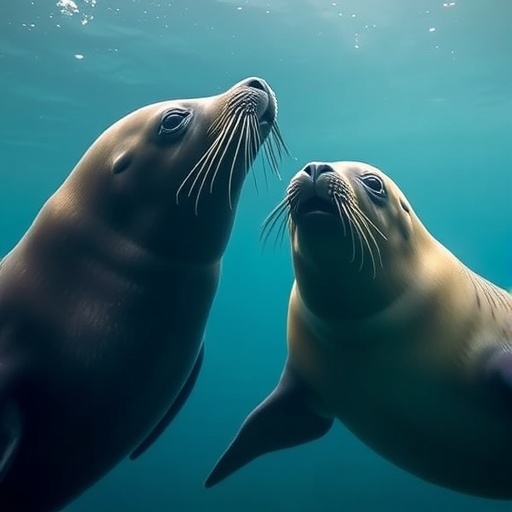
The study undertook a comparative approach, analyzing the neural architecture of a range of species including coyotes, California sea lions, harbor seals, and northern elephant seals. These marine carnivores, all bearing close evolutionary ties to canines, exhibit varying degrees of vocal control. Seals distinguish themselves as one of the few animal groups capable of impressive vocal plasticity, even mimicking human sounds—a trait seldom found outside of humans and some bird species. Sea lions demonstrate a more modest, yet notable, capacity for vocal flexibility.
Lead researchers Gregory Berns, a psychology professor at Emory, and Peter Cook, now a marine mammal science professor at New College of Florida, leveraged advanced diffusion magnetic resonance imaging (MRI) techniques to scrutinize the white matter pathways of post-mortem brains. Diffusion MRI is a specialized form of imaging that tracks the movement of water molecules through brain tissue, revealing connectivity patterns between regions otherwise invisible through conventional methods. This non-invasive imaging allows an unprecedented view into the wiring diagrams of animal brains, offering granular insights into evolutionary neurobiology.
The origin of the brains studied is equally compelling—all specimens were sourced ethically from wild animals that either died naturally or were humanely euthanized due to injury in rehabilitation settings. This approach preserves biological authenticity while permitting detailed neuroscientific investigation beyond the constraints of live-subject imaging.
The central discovery of the research lies in the identification of a neural “bypass” circuit within the marine mammal brains. Unlike the coyote brains, where vocalization is governed through the mid-brain—a hub associated with automatic, survival-related functions such as breathing and threat response—the sea lions and seals possess a direct monosynaptic connection from the vocal motor cortex to the brainstem neurons controlling the larynx muscles. This anatomical bypass effectively circumvents the mid-brain, granting these marine mammals conscious, voluntary command over their vocal output.
This neural architecture is hypothesized to underpin the vocally flexible behaviors seen in these species. In contrast, most other mammals’ vocalizations remain inflexible due to the obligatory engagement of mid-brain circuits, which constrains call variability and prevents complex vocal learning. By decoupling vocal control from automatic mid-brain pathways, seals and sea lions have unlocked an evolutionary niche for vocal plasticity.
One driver behind this adaptation is the demanding respiratory control required for underwater foraging. Pinnipeds—seals, sea lions, and elephant seals—have evolved sophisticated breathing and swallowing strategies to accommodate prolonged dives and underwater feeding. For example, sea lions can remain submerged for up to 20 minutes, while some elephant seal species can extend this duration to nearly two hours, necessitating exceptional voluntary breath control. The researchers posit that these ecological pressures inadvertently sculpted vocal control pathways in the brain, producing the neural foundation for learning-controlled vocalization.
This study also draws attention to the evolutionary broader implications of vocal learning and the neurobiological prerequisites that facilitate it. Peter Cook eloquently summarizes, “We’ve uncovered an ecological recipe for how a mammal might evolve a vocally flexible brain.” This finding not only advances understanding of pinniped communication but also offers a tantalizing framework for reconstructing the evolutionary tree leading to human language.
By extending this comparative imaging approach across a diverse set of mammalian species, the researchers aspire to map the progressive stages of vocal control evolution. Gregory Berns envisions that such neuroimaging studies could eventually inform the deep evolutionary origins of speech by establishing connectivity patterns that correlate with vocal learning capacities.
The technical prowess of diffusion MRI on preserved brains owes its development to Karla Miller of the University of Oxford, who initially applied this method to probe Alzheimer’s pathology in human post-mortem brain tissue. The fixative properties of formaldehyde preservation eliminate motion artifacts, enabling exceptionally high-resolution imaging over prolonged scan durations—a clear advantage over live imaging constraints.
Berns and his collaborators have pioneered applying this technique to a vast array of animals, including historical brain specimens from museum collections. Their 2017 work mapping neural connectivity in thylacine brains preserved for over a century demonstrates the robustness and longevity of diffusion MRI data, vastly extending the temporal reach of neurobiological research.
The study also nuances our perspectives of pinnipeds’ cognitive abilities. Despite common perceptions of seals and sea lions as slow, lumbering creatures, Cook highlights their intellectual acumen, noting brain sizes comparable to chimpanzees and pronounced motivation for learning auditory and memory tasks. This cognitive sophistication complements their neuroanatomical adaptations for vocal control, making them fascinating models for studying the biological underpinnings of communication.
Intriguingly, the data reveal species-specific differences in neuroanatomical pathways related to auditory-vocal integration. Harbor seals, for instance, demonstrate particularly pronounced connectivity between the thalamus—a crucial sensorimotor relay center—and the vocal motor cortex. This circuit mirrors structures in parrots and humans known to facilitate vocal learning, suggesting convergent evolution of neural substrates that support mimicry and complex sound production.
Future research avenues include expanding these investigations into other marine mammals such as whales, dolphins, and porpoises, which exhibit exceptional vocal abilities. Such work promises to deepen our grasp of the convergent evolutionary processes that foster vocal learning across distantly related mammalian lineages.
Peter Cook aptly captures the essence of this field of study by stating, “All animals can learn, and almost all birds and mammals communicate with their voices. The paradox of why so few animals can learn to control their calls remains an irresistible scientific mystery.” This research brings us closer to solving that puzzle by revealing the distinct neurobiological infrastructure required for vocal learning, highlighting the intricate intersections of ecology, neuroanatomy, and evolutionary biology.
Subject of Research: Animals
Article Title: Seal and Sea Lion Brains Have Evolved to Support Volitional Control of Vocal Behavior and Learning
News Publication Date: 12-Mar-2026
Web References: https://dx.doi.org/10.1126/science.adx9367
Image Credits: C. Reichmuth
Tags: brain evolution and speechcomparative neuroanatomy of carnivoresdiffusion MRI in neuroscience researchevolution of vocalization in marine mammalsevolutionary basis of speech in animalsmarine mammal communication studiesmimicry of human sounds by sealsneural adaptations for vocal controlneurobiology of vocal learningvocal flexibility in seals and sea lionsvocal plasticity in marine mammalsvoluntary breathing control in aquatic animals

