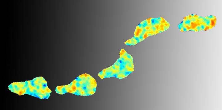

Credit: Pontus Nordenfelt
It's a known fact that cells can move around the body, but how they do it has been unknown – until now. Researcher in Infection Medicine Pontus Nordenfelt at Lund University in Sweden has managed to describe and visualize cell migration on a molecular level. In time, this could become significant in the treatment of infectious diseases, inflammation, cancer, etc. where cell migration plays an important role.
In his studies, Pontus Nordenfelt used T cells as models. The T cells are part of the immune system and must therefore be able to reach tissue which has been exposed to, for instance, a bacterial invasion.
To be able to move, the cell must attach itself to a surface and use its front to push to exert the force it needs. Meanwhile, the rear part of the cell must let go from the surface, allowing it to "roll" forward, so to speak.
"When moving, the cell converts chemical energy into mechanical force. This force can now be measured using the microscope tool I developed", says Pontus Nordenfelt, an engineer from the start, whose work benefited greatly from having this background.
Cell migration is enabled by three molecules working together. Integrins are molecules located on the cell surface that are able to attach themselves to other surfaces. Actin are small building blocks on the inside of the cell membrane, and collectively form the skeleton of the cell. Finally, the "adaptor" is a protein that links integrin and actin together.
"Once the adaptor has connected the integrins and actin, the mechanical force from actin gives the 'green light'. The integrin molecule then binds itself to a nearby partner surface, through which the cell can move slightly forward. Once the cell's migration is complete, the integrin and actin molecules separate from each other in this part of the cell, while another part of the cell becomes active", explains Pontus Nordenfelt.
The key to cell migration is that the integrin molecules become active in the appropriate part of the cell, and how this works has so far been unclear. The Lund researcher's study is published in Nature Communications.
The notion that cell migration would be based on activation of integrins through force from actin was suggested several years ago by prominent researcher Timothy Springer at Harvard, where Pontus Nordenfelt spent three years examining how integrins become active. Among other things, the work resulted in new microscopy techniques that have been used to test this hypothesis of how cell migration works.
The technique is based on fluorescent biosensors that are inserted into the cell, showing force changes in integrins through variations in colour. This allows you to see the process of cell migration through colour images, and measure the mechanical force acting on the integrins. This technique could enable the development of more targeted drugs to strengthen the body's immune system against infections, or prevent tumours from metastasising, etc.
As a researcher in Infection Medicine, Pontus Nordenfelt became interested in integrins, as this is a group of proteins used by the immune cells, which led to his studies of cell migration. With the help of his new tool, he now wants to continue with the study of phagocytes – immune cells that "eat" invading bacteria.
"The fluorescent biosensors enable measuring when and how different bacteria are ingested; perhaps it's also possible to create variants of the sensors that measure the opposite, that is, when bacteria avoid being eaten and instead manage to invade the body", he says.
###
The research article published in Nature Communications: Coordinated integrin activation by actin-dependent force during T cell migration http://www.nature.com/articles/ncomms13119
Pontus Nordenfelt can be reached at tel +46 (0)46 222 85 71/+46 (0)731 523 553 or email [email protected] For more information, see also http://nordlab.med.lu.se
Pontus Nordenfelt is a postdoc researcher and part of the Lund Faculty of Medicine, a creative research environment known as SEBRA (Sepsis and Bacterial Resistance Alliance). He is also chair of the collective network of junior medicine researchers in Lund, Future Faculty.
VIDEOS
1, Cells in normal movement
https://www.youtube.com/watch?v=ORBSW7G92Nc&feature=youtu.be
Cells in normal movement on a glass surface, filmed using light microscopy.
2, Cells that are unable to move properly
https://www.youtube.com/watch?v=Klxo4-dyWw8&feature=youtu.be
Cells that are unable to move properly due to failure of the integrin link. Filmed using light microscopy.
3, Cell with an integrin sensor filmed using multichannel fluorescence microscopy
https://www.youtube.com/watch?v=zje2XrlpE2M&feature=youtu.be
The film shows a cell with an integrin sensor, filmed using multichannel fluorescence microscopy. Automatised computer-based analysis has divided the cells into boxes depending on their shape and speed. This is to enable subsequent comparison of equivalent areas between different cells. The force exerted on the integrin is shown on a blue-red scale where red is strongest.
4, Force changes in integrins https://www.youtube.com/watch?v=aXhDXHaVXZk&feature=youtu.be
The film shows cells with integrin sensors filmed using multichannel fluorescence microscopy. The force exerted on the integrin is shown on a blue-red scale where red is strongest.
Media Contact
Pontus Nordenfelt
[email protected]
46-731-523-553
@lunduniversity
http://www.lu.se

