
Credit: Peter Morenus/UConn Photo
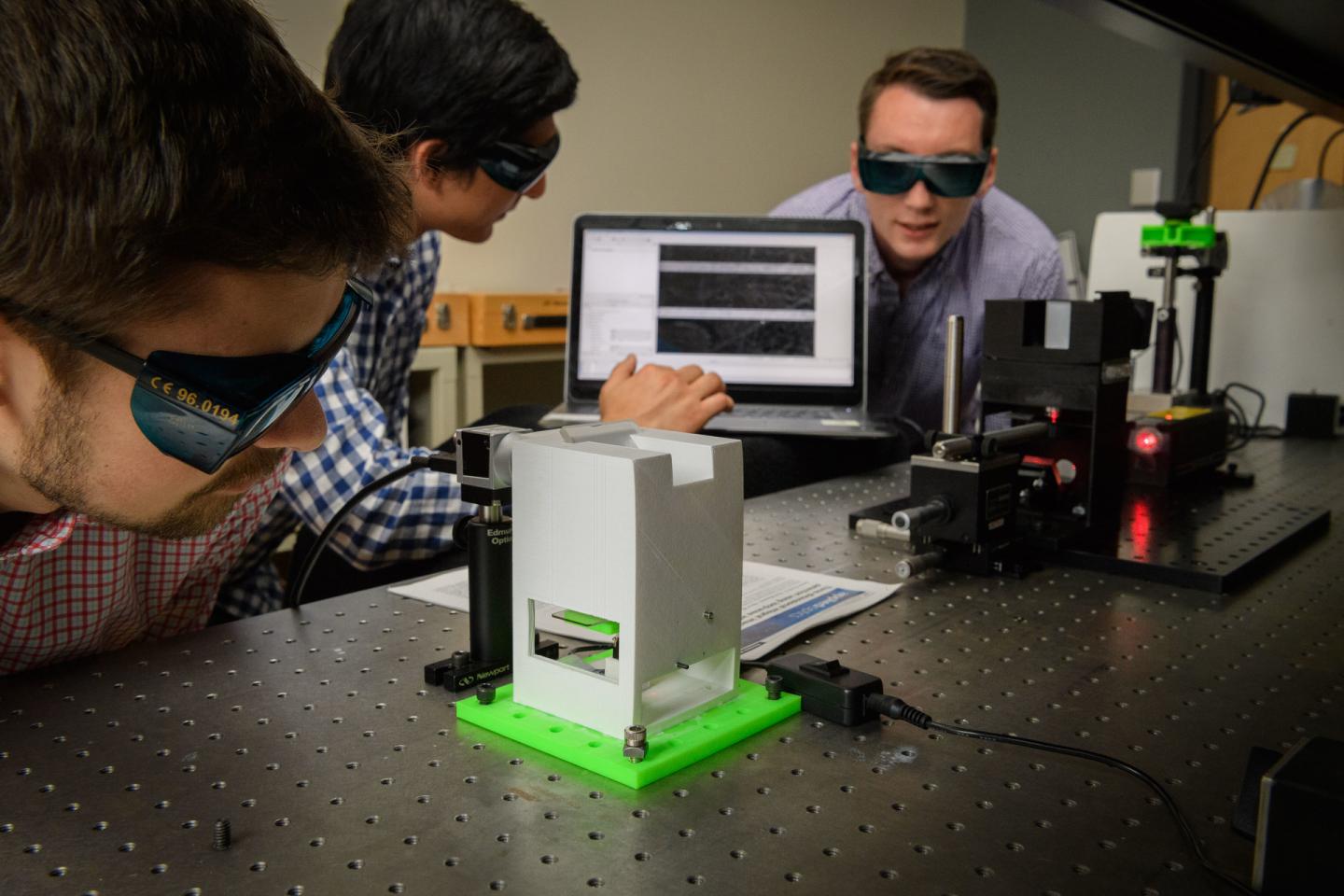
A portable holographic field microscope developed by University of Connecticut optical engineers could provide medical professionals with a fast and reliable new tool for the identification of diseased cells and other biological specimens.
The device, featured in a recent paper published by Applied Optics, uses the latest in digital camera sensor technology, advanced optical engineering, computational algorithms, and statistical analysis to provide rapid automated identification of diseased cells.
One potential field application for the microscope is helping medical workers identify patients with malaria in remote areas of Africa and Asia where the disease is endemic.
Quick and accurate detection of malaria is critical when it comes to treating patients and preventing outbreaks of the mosquito-borne disease, which infected more than 200 million people worldwide in 2015, according to the Centers for Disease Control. Laboratory analysis of a blood sample remains the gold standard for confirming a malaria diagnosis. Yet access to trained technicians and necessary equipment can be difficult and unreliable in those regions.
The microscope's potential applications go far beyond the field diagnosis of malaria. The detailed holograms generated by the instrument also can be used in hospitals and other clinical settings for rapid analysis of cell morphology and cell physiology associated with cancer, hepatitis, HIV, sickle cell disease, heart disease, and other illnesses, the developers say.
In checking for the presence of disease, most hospitals currently rely on dedicated laboratories that conduct various tests for cell analysis and identification. But that approach is time consuming, expensive, and labor intensive. It also has to be done by skilled technicians working with the right equipment.
"Our optical instrument cuts down the time it takes to process this information from days to minutes," says Bahram Javidi, Board of Trustees Distinguished Professor in the Department of Electrical and Computer Engineering and the microscope's senior developer. "And people running the tests don't have to be experts, because the algorithms will determine if a result is positive or negative."
The research team consulted with hematologists, and the algorithms used with the instrument are able to compare a sample against the known features of healthy cells and the known features of diseased cells in order to make proper identification. "It's all done very quickly," Javidi says.
How the Device Works
When it comes to identifying patients with malaria, here's how the device works: A thin smear from a patient's blood sample is placed on a glass side, which is put under the microscope for analysis. The sample is exposed to a monochromatic light beam generated by a laser diode or other light source. Special components and optical technologies inside the microscope split the light beam into two beams in order to record a digital hologram of the red blood cells in the sample. An image sensor, such as a digital webcam or cell phone camera, connected to the 3-D microscope captures the hologram. From there, the captured data can be transferred to a laptop computer or offsite laboratory database via the internet. Loaded with dedicated algorithms, the computer or mobile device hardware reconstructs a 3-D profile of the cell and measures the interaction of light with the cell under inspection. Any diseased cells are identified using computer pattern recognition software and statistical analysis.
Red blood cells infected with the malaria-causing Plasmodium parasite exhibit different properties than healthy blood cells when light passes through them, Javidi says.
"Light behaves differently when it passes through a healthy cell compared to when it passes through a diseased cell," Javidi says. "Today's advanced sensors can detect those subtle differences, and it is those nanoscale variations that we are able to measure with this microscope."
Conventional light microscopes only record the projected image intensity of an object, and have limited capability for visualizing the detailed quantitative characterizations of cells. The digital holograms acquired by UConn's 3-D microscope, on the other hand, capture unique micro and nanoscale structural features of individual cells with great detail and clarity. Those enhanced images allow medical professionals and researchers to measure an individual cell's thickness, volume, surface, and dry mass, as well as other structural and physiological changes in a cell or groups of cells over time – all of which can assist in disease identification, treatment, and research. For instance, the device could help researchers see whether new drugs impact cells positively or negatively during clinical trials.
The techniques associated with the holographic microscope also are non-invasive, highlighting its potential use for long-term quantitative analysis of living cells.
Conventional methods of testing blood samples for disease frequently involve labeling, which means the sample is treated with a chemical agent to assist with identification. In the case of malaria, red blood cells are usually treated with a Giemsa stain that reacts to proteins produced by malaria-carrying parasites and thus identifies them. But introducing a chemical into a live cell can change its behavior or damage it.
"If you're doing an in vitro inspection of stem cells, for instance, and you introduce a chemical agent, you risk damaging those cells. And you can't do that, because you may want to introduce those cells into the human body at some point," Javidi says. "Our instrument doesn't rely on labeling, and therefore avoids that problem."
###
The holographic microscope was developed in UConn's new Multidimensional Optical Sensing & Imaging Systems or MOSIS lab, where Javidi serves as director. The MOSIS lab integrates optics, photonics, and computational algorithms and systems to advance the science and engineering of imaging from nano to macro scales.
A comprehensive report on the MOSIS lab's work with 3-D optical imaging for medical diagnostics was published last year in Proccedings of the IEEE, the top-ranked journal for electrical and electronics engineering. Joining Javidi in this research are graduate students Adam Markman, Siddharth Rawat, Satoru Komatsu, and Tim O'Connor from UConn; and Arun Anand, an applied optics specialist with Maharaja Sayajirao University of Baroda in Vadodara, India.
The microscope research is supported by Nikon and the National Science Foundation (ECCS 1545687). Students are supported by the U.S. Department of Education, GE, and Canon fellowships. Other sponsors that have supported Javidi's broader research work and the MOSIS lab over the years include the Defense Advanced Research Projects Agency or DARPA, the U.S. Airforce Research Lab, the U.S. Army, the Office of Naval Research, Samsung, Honeywell, and Lockheed Martin. He has collaborated with colleagues from numerous universities and industries around the world during his time at UConn, including research facilities in Japan, Korea, China, India, Germany, England, Italy, Switzerland, and Spain, among other countries.
Javidi is working with colleagues at UConn Health, including medical oncology and hematology specialist Dr. Biree Andemariam and her staff, for other medical applications. UConn's tech commercialization office has been involved in discussing potential marketing opportunities for the portable digital microscope.
A prototype of the microscope used for initial tests was assembled using 3-D printing technologies, lowering its production costs.
Media Contact
Colin Poitras
[email protected]
860-486-4656
@UCToday
Home
Original Source
https://today.uconn.edu/2017/10/giving-cell-disease-closer-look/ http://dx.doi.org/10.1364/AO.56.00D127


