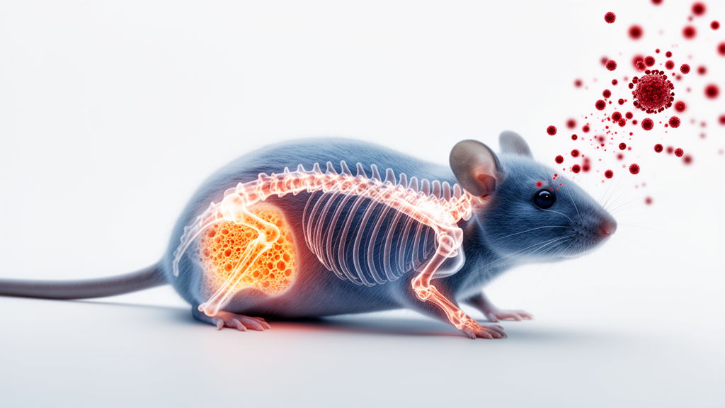
Mice engineered to carry HIV genes but never actually infected with the virus lose bone at a young age, according to a new study published in Frontiers in Endocrinology. The finding, from researchers at the University of Barcelona and collaborators, suggests that HIV proteins themselves—rather than antiretroviral drugs or uncontrolled viral replication—can be sufficient to disrupt the delicate balance of bone formation and resorption that keeps the skeleton intact.
People living with HIV are known to face elevated risks of reduced bone mineral density, osteopenia, osteoporosis, and fragility fractures compared with HIV-negative populations, even when they receive suppressive antiretroviral therapy. Disentangling the causes has proven difficult in human studies, because viral infection, drug toxicity, chronic inflammation, lifestyle factors, and comorbidities such as hepatitis coinfection all converge on the skeleton. The Barcelona team turned to an animal model to isolate one of these variables: the direct biological activity of HIV-derived proteins.
The researchers used the heterozygous Tg26 mouse, a well-established HIV-1 transgenic strain that constitutively expresses multiple HIV-1 viral proteins without productive viral replication. Male Tg26 mice and wild-type littermates were maintained for either 6 or 25 weeks, corresponding to young and adult animals, before blood and tissues were collected. The team confirmed active viral transcription by measuring Tat mRNA, which was substantially expressed across all tissues analyzed, particularly in white adipose tissue depots and in bone itself.
Before examining the skeleton, the researchers characterized the metabolic and inflammatory state of the animals. Young Tg26 mice weighed less than controls, and this deficit persisted into adulthood. The reduction was driven by adipose tissue: both subcutaneous inguinal and visceral epididymal fat depots were significantly smaller in transgenic mice at both ages, while liver mass, tibia length, and food intake remained unchanged. Glucose and insulin levels were unaltered, indicating preserved glucose homeostasis despite the marked lipoatrophy. Serum leptin, produced by fat tissue, was dramatically reduced at both ages, consistent with the smaller fat depots.
Inflammation showed only a mild profile. Tumor necrosis factor-alpha was significantly elevated in 6-week-old transgenic mice, and interleukin-6 and plasminogen activator inhibitor-1 showed a tendency to increase at 25 weeks, but the overall systemic inflammatory changes were modest. This matters for the bone findings, because chronic inflammation is one of the proposed mechanisms linking HIV to skeletal disease, and the Tg26 model displays only a low-grade inflammatory signature rather than the intense immune activation seen in uncontrolled infection.
The skeletal analysis centered on high-resolution micro-computed tomography of fixed femurs, performed according to American Society of Bone and Mineral Research guidelines. Multiple cortical and trabecular parameters were significantly reduced in transgenic mice. Cortical bone volume and thickness decreased in both young and older Tg26 animals, although the cross-sectional perimeter of the femur was preserved, indicating that overall skeletal development proceeded normally even as bone acquisition and maintenance faltered. Trabecular bone, the spongy internal network, showed reduced thickness and number and increased separation, and the bone volume fraction—the proportion of mineralized bone per unit volume—was also lower in transgenic mice.
Molecular analysis of calvarial bone pointed to a remodeling system tilted away from bone formation. Expression of Rankl, the ligand that drives osteoclast differentiation and activity, and of Sost, the gene encoding sclerostin, a potent inhibitor of the Wnt/beta-catenin pathway essential for osteoblast differentiation, were both elevated, especially in the older mice. Runx2, a master regulator of early osteoblast differentiation, showed a significant genotype-by-age interaction, as did Dkk1 and periostin, indicating dysregulated remodeling pathways. Matrix metalloproteinase-13, involved in extracellular matrix turnover, was reduced. Notably, several other markers—Fgf23, Col1a1, osteopontin (Spp1), and Osterix (Sp7)—showed no changes, suggesting that core osteoblast activity, matrix production, and phosphate homeostasis were largely preserved.
Circulating bone turnover markers reinforced the picture of a high-turnover state. CTX-1, a marker of osteoclast-mediated bone resorption, was significantly elevated in transgenic mice, and P1NP, a marker of osteoblast activity and bone formation, showed a tendency to increase in the 25-week-old animals. Together, these changes indicate coupled high-turnover bone remodeling, in which both resorption and formation accelerate but net bone is lost. Circulating osteoprotegerin, an anti-osteoclastogenic factor that inhibits RANKL signaling, was also elevated in the older transgenic mice; the authors interpret this as a possible compensatory counter-regulatory response rather than a primary driver of the phenotype.
The authors acknowledge important limitations. The study included only male animals, so potential sex-dependent effects could not be assessed. The sample size in older mice was relatively small, limiting statistical power. The transcriptional changes were measured in calvarial bone while the morphometric phenotype was assessed in the femur, so confirming the molecular alterations at the anatomical site of bone loss remains an open task. The heterozygous transgenic model also expresses a defined set of viral proteins constitutively and does not fully recapitulate natural infection, with its dynamic viral replication and broader repertoire of viral factors and host immune responses.
One alternative explanation the researchers specifically addressed was kidney disease. The Tg26 model is known to develop renal pathology, and bone abnormalities could in principle be secondary to renal mineral disturbances. However, because the FGF23–kidney axis is central to phosphate and mineral homeostasis, a purely renal mechanism would be expected to alter FGF23. Both Fgf23 mRNA expression in calvaria and circulating FGF23 levels remained unchanged, and the selective pattern of altered genes—sclerostin, matrix metalloproteinase-13, and periostin changing while Sp7, Spp1, and Col1a1 were preserved—supports a model in which bone remodeling is specifically perturbed rather than globally disrupted by kidney failure. Within these constraints, the study strengthens the argument that HIV-associated skeletal disease is not exclusively a consequence of antiretroviral treatment, and that viral proteins acting directly on bone cells and the bone microenvironment may contribute meaningfully to the bone loss seen in people living with HIV.
Subject of Research: The effects of HIV viral protein expression on bone homeostasis in a transgenic mouse model
Article Title: HIV viral protein expression disrupts bone tissue homeostasis in mice
Article References: Villarroya, J., Artigas, N., Quesada-López, T., Cereijo, R., Domingo, P., Villarroya, F., Ventura, F., & Giralt, M. (2026). HIV viral protein expression disrupts bone tissue homeostasis in mice. Frontiers in Endocrinology, 17, Article 1920538. https://doi.org/10.3389/fendo.2026.1920538
Image Credits: AI Generated
DOI: 10.3389/fendo.2026.1920538
Keywords: HIV, bone loss, osteoporosis, Tg26 transgenic mice, bone remodeling, osteoclasts, osteoblasts, RANKL, microCT, lipoatrophy, bone turnover markers, antiretroviral therapy
News Source: Kristina Jarvis. (October 27, 2026). HIV Viral Proteins Alone Drive Bone Loss in Transgenic Mice. Scienmag.