In an era where the complexity of biological systems demands increasingly sophisticated analytical tools, a groundbreaking advancement promises to revolutionize spatial transcriptomics. Traditional fluorescence microscopy, while fundamental to biological imaging, has long been hamstrung by its limited multiplexing capacity, typically allowing the simultaneous visualization of only a handful of RNA targets. This constraint has posed a significant obstacle for researchers aiming to map the intricate spatial distributions of numerous RNA species within cells and tissues. However, a newly introduced method termed “profiling of RNA in situ through single-round imaging” (PRISM) transcends these limitations, enabling unprecedented high-plex RNA imaging in a single round with conventional microscopes.
The marvel of PRISM lies in its ingenious use of color intensity grading to expand the coding capacity of spatial RNA imaging, obviating the need for cumbersome cyclic reactions prevalent in earlier multiplexing techniques like sequential fluorescence in situ hybridization (seqFISH). While seqFISH and similar approaches rely on multiple rounds of hybridization and imaging to achieve multiplexing, PRISM accomplishes this feat in just one imaging cycle. This not only simplifies the experimental workflow but also eliminates the requirement for specialized instrumentation, thereby democratizing access to high-plex spatial transcriptomics.
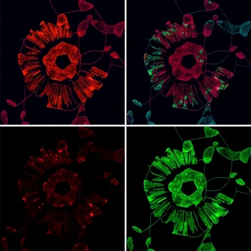
At the core of PRISM’s innovation is a radius vector filtering strategy, a computational approach designed to maintain the distinguishability of color barcodes in a multidimensional color space. By carefully optimizing the spacing between color intensity codes, PRISM ensures that each RNA species can be accurately identified without signal overlap or misclassification, a challenge that has historically limited multiplexing capacities in fluorescence imaging. This methodological refinement allows PRISM to achieve up to 64-plex barcoded RNA imaging within a single round, a staggering leap forward from conventional capabilities.
The implications of such multiplexing power are vast. PRISM was rigorously validated across diverse biological contexts, showcasing its versatility and robustness. One notable demonstration involved the construction of a detailed three-dimensional atlas of mouse embryonic development spanning from embryonic day 12.5 to 14.5. This temporal and spatial mapping of RNA expression profiles provides unprecedented insights into the orchestrated molecular dynamics driving embryogenesis, capturing cellular heterogeneity and tissue differentiation with remarkable precision.
Beyond developmental biology, PRISM’s potential was further illustrated through its application to pathological specimens. The technology enabled the generation of a quasi-three-dimensional landscape of tumor-normal transitions in human hepatocellular carcinoma tissues. This spatially resolved molecular cartography unveiled complex interplays within the tumor microenvironment, highlighting heterogeneity and distinct cellular neighborhoods that contribute to cancer progression and therapeutic resistance.
Moreover, in the realm of neuroscience, PRISM facilitated the assembly of a comprehensive 3D cell atlas and detailed subcellular RNA localization maps of the mouse brain. Such fine-scale imaging elucidates the spatial distribution of transcripts at the subcellular level, providing critical information on neuronal identity, function, and intercellular communication pathways integral to brain physiology and disease mechanisms.
One of the most compelling findings leveraging PRISM was the revelation of the critical role played by cancer-associated fibroblasts (CAFs) in modulating immune cell infiltration and immune response heterogeneity within tumor microenvironments. By spatially profiling RNA expression patterns of both stromal and immune components, PRISM illuminated how CAFs orchestrate immunological niches that influence tumor behavior and potentially impact therapy outcomes. This insight opens new avenues for designing targeted immunomodulatory treatments in oncology.
Unlike previous multiplexing approaches that require multiple rounds of probe hybridization and imaging—often demanding advanced automation and protracted experimental timelines—PRISM streamlines the spatial transcriptomics workflow. Single-round imaging reduces photobleaching, sample distortion, and cumulative errors, thereby enhancing data reliability and reproducibility. Its compatibility with existing fluorescence microscopy platforms accelerates adoption and facilitates integration into ongoing research projects.
The architecture of PRISM hinges on precise control and quantification of fluorescence signal intensities, translating subtle gradations in color channels into discrete coded barcodes. This strategy surmounts the spectral overlap limitations inherent in traditional fluorophore-based detection systems, enabling denser packing of information without expanding spectral bands. Consequently, researchers can now spatially resolve dozens of RNA species simultaneously, dramatically amplifying throughput and data richness.
Furthermore, the computational sophistication embedded in PRISM’s color decoding algorithms is a crucial enabler of its success. By employing radius vector filtering to define minimum distances between color code clusters, the system mitigates false positives and enhances quantitative accuracy. This advancement exemplifies the growing convergence of computational innovation and molecular imaging in modern biosciences.
PRISM’s capacity for high-throughput, multiplexed spatial RNA profiling is poised to impact a broad spectrum of biological disciplines, from developmental and cancer biology to neuroscience and immunology. The ability to visualize extensive RNA landscapes in situ within intact tissues unlocks new dimensions of understanding in cellular architecture, molecular interactions, and dynamic biological processes.
Looking ahead, the adoption of PRISM may catalyze the development of integrated platforms combining spatial transcriptomics with other omics modalities, fostering comprehensive multi-dimensional cellular profiling. Its compatibility with conventional microscopes highlights a trend toward making advanced molecular imaging accessible and scalable, potentially accelerating translational research and clinical diagnostics.
In sum, PRISM represents a paradigm shift in spatial RNA imaging, breaking through previous multiplexing barriers with a clever fusion of color intensity coding and computational filtering. By marrying simplicity with sophistication, it empowers researchers to decode the complex spatial RNA landscapes of health and disease with unprecedented clarity – all within the familiar confines of single-round, conventional microscopy.
As this technology gains traction, the life sciences community stands on the cusp of a new era where expansive, high-resolution spatial transcriptomic maps will become routine tools, propelling discoveries in fundamental biology and precision medicine alike. PRISM’s innovative approach is a testament to the power of creative problem-solving at the intersection of imaging science and molecular biology, charting a bold path forward for the future of spatial genomics.
Subject of Research: Spatial transcriptomics; RNA imaging; multiplexed fluorescence microscopy; tumor microenvironment; embryonic development; neuroscience.
Article Title: High-plex spatial RNA imaging in one round with conventional microscopes using color-intensity barcodes.
Article References:
Chang, T., Zhao, S., Deng, K. et al. High-plex spatial RNA imaging in one round with conventional microscopes using color-intensity barcodes. Nat Biotechnol (2025). https://doi.org/10.1038/s41587-025-02883-7
Image Credits: AI Generated
Tags: advanced fluorescence microscopy techniquescolor-intensity barcoding in microscopydemocratization of spatial transcriptomicshigh-plex spatial RNA imaginginnovative analytical tools in biologymultiplexing in RNA visualizationovercoming limitations of seqFISHPRISM method in transcriptomicsRNA hybridization methodssimplified experimental workflowssingle-round imaging techniquesspatial distribution of RNA species

