


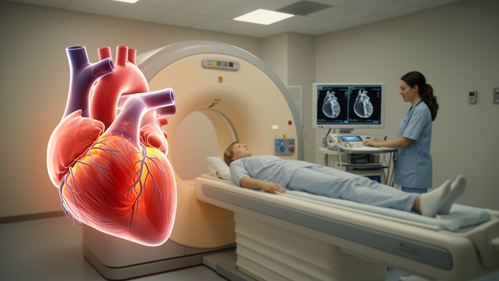
Patients recovering from a heart attack may not need contrast dye for the MRI scans that guide their follow-up care, according to a new retrospective study published in BMC Medical Imaging. Combining two contrast-free imaging measurements — left ventricular global longitudinal strain (LV GLS) and native T1 mapping — predicted major adverse cardiac events after acute myocardial infarction at least as well as, and in some comparisons better than, conventional contrast-enhanced markers.
In the retrospective study, 1253 patients from four centers who underwent percutaneous coronary intervention and cardiac MRI between February 2017 and September 2024 were enrolled. The cohort was split into a development group of 1002 patients and an external validation group of 251. Over a median follow-up of 43 months, 137 patients in the development cohort and 36 in the validation cohort experienced a major adverse cardiac event, defined as all-cause death, life-threatening arrhythmias, cardiac death, reinfarction or heart failure hospitalization.
The key comparison centered on two risk models. Model 2 relied on conventional contrast-enhanced CMR markers: late gadolinium enhancement, microvascular obstruction and intramyocardial hemorrhage. Model 3 instead combined clinical variables, left ventricular ejection fraction, native T1 z score and LV GLS, all obtainable without contrast. In the development cohort, the non-contrast model improved discrimination over the contrast-enhanced model, with a C-index of 0.780 versus 0.736 (P = 0.033). The pattern held in the external validation cohort.
The study found that these two measures together offered complementary prognostic information, improving risk stratification and reclassification beyond established risk factors.
The non-contrast approach also outperformed two established clinical tools. Native T1 and LV GLS showed better prognostic performance than both the GRACE score and the Glasgow CMR Risk Score, suggesting that the imaging-based combination captures prognostic information these scores miss.
The authors conclude that combined native T1 mapping and LV GLS supported the potential of a contrast-free CMR strategy that outperformed a conventional contrast-enhanced model. Because the study was retrospective and focused on AMI patients who had already undergone PCI and CMR, the findings describe prognostic associations in this population rather than the effect of changing clinical practice.
Subject of Research: Prognostic value of non-contrast cardiac MRI markers after acute myocardial infarction
Article Title: Prognosis and risk stratification with LV global longitudinal strain and Native T1 mapping in acute myocardial infarction
Article References: Zhang, S., Zhu, G., Gu, X., Wu, J., & Wu, L. (2026). Prognosis and risk stratification with LV global longitudinal strain and Native T1 mapping in acute myocardial infarction. BMC Medical Imaging. https://doi.org/10.1186/s12880-026-02992-y
Image Credits: AI Generated
DOI: 10.1186/s12880-026-02992-y
Keywords: cardiac MRI, acute myocardial infarction, native T1 mapping, global longitudinal strain, risk stratification, major adverse cardiac events, prognosis, contrast-free imaging, late gadolinium enhancement, C-index, risk, stratification
News Source: Ophelia Keating. (October 28, 2026). Heart MRI Without Contrast Dye Predicts Risk After Heart Attack. Scienmag.



