• HOME
  • NEWS
  • EXPLORE
    • CAREER
      • Companies
      • Jobs
    • EVENTS
    • iGEM
      • News
      • Team
    • PHOTOS
    • VIDEO
    • WIKI
  • BLOG
  • COMMUNITY
    • FACEBOOK
    • INSTAGRAM
    • TWITTER
Sunday, August 10, 2025
BIOENGINEER.ORG
No Result
View All Result
  • Login
  • HOME
  • NEWS
  • EXPLORE
    • CAREER
      • Companies
      • Jobs
        • Lecturer
        • PhD Studentship
        • Postdoc
        • Research Assistant
    • EVENTS
    • iGEM
      • News
      • Team
    • PHOTOS
    • VIDEO
    • WIKI
  • BLOG
  • COMMUNITY
    • FACEBOOK
    • INSTAGRAM
    • TWITTER
No Result
View All Result
Bioengineer.org
No Result
View All Result
Home NEWS Science News Stem Cells

Growing brains in the lab

Bioengineer by Bioengineer
February 8, 2014
in Stem Cells
Reading Time: 2 mins read
0
Share on FacebookShare on TwitterShare on LinkedinShare on RedditShare on Telegram

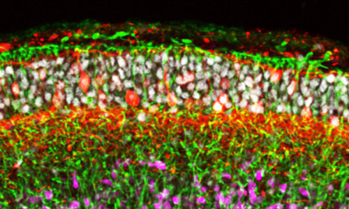
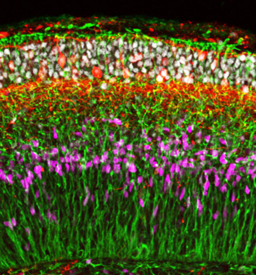
Human embryonic stem cells can be induced to spontaneously form developing brain tissue During development, the nervous system forms as a flat sheet called the neuroepithelium on the outer layer of the embryo. This sheet eventually folds in on itself to form a neural tube that gives rise to the brain and spinal cord—a process that involves the proliferation and migration of immature nerve cells to form the brain at one end and the spinal cord at the other. Yoshiki Sasai, Taisuke Kadoshima and colleagues from the RIKEN Center for Developmental Biology have now shown that human embryonic stem (ES) cells can spontaneously organize into the cerebral cortical tissue that forms at the front, or ‘brain’ end, of the developing neural tube1.

Figure: Human embryonic stem cells spontaneously organize into neuroepithelial tissue containing multiple zones after growing for 70 days in culture. © 2014 Yoshiki Sasai, RIKEN Center for Developmental Biology
Figure: Human embryonic stem cells spontaneously organize into neuroepithelial tissue containing multiple zones after growing for 70 days in culture.

Sasai and his colleagues previously developed a novel cell culture technique that involves growing ES cells in suspension, and have shown that these cells can self-organize into complex three-dimensional structures. They have already used this method to grow pieces of cerebral cortex and embryonic eyes from mouse ES cells. And more recently, they have shown that human ES cells can also organize into embryonic eyes containing retinal tissue and light-sensitive cells.

In their most recent work, Sasai’s team treated human ES cells grown using their cell culture system with signaling molecules that induce the formation of nervous tissue from the outer embryonic layer. They found that the cells spontaneously organize into neuroepithelial tissue that then folds up to give a multilayered cortex (Fig. 1).

During human embryonic development, the neural tube thickens at both ends. In particular, the front end thickens dramatically as waves of cells migrate outward to form the layered cerebral cortex and other parts of the brain. An important finding of the team’s is that the front end of the neural tube appears to thicken due to the growth of radial glial fibers, which span the thickness of the tube and guide migrating cells, rather than due to the accumulation of immature cells within the tube, as previously thought.

The findings also highlight critical differences between the development of the neural tube in mice and humans. While in humans, the inner surface of the neural tube and the intermediate neuroepithelial zone underneath it contain distinct populations of neural progenitors resembling radial glia, the progenitor population in the latter is not present in the developing mouse cortex.

“Efficient generation of cortical tissues could provide a valuable resource of functional neurons and tissues for medical applications,” says Kadoshima. “By combining this method with disease-specific human induced pluripotent stem cells, it will also be possible to reproduce complex human disorders.”

Story Source:

The above story is based on materials provided by RIKEN Center for Developmental Biology.
Photo: © 2014 Yoshiki Sasai, RIKEN Center for Developmental Biology

Share13Tweet8Share2ShareShareShare2

Related Posts

blank

Human stem cells treat spinal cord injury side effects in mice

October 4, 2016
blank

Research into fly development provides insights into blood vessel formation

September 30, 2016

Fertility genes required for sperm stem cells

September 28, 2016

Regulatory RNA essential to DNA damage response

September 27, 2016
Please login to join discussion

POPULAR NEWS

  • blank

    Molecules in Focus: Capturing the Timeless Dance of Particles

    138 shares
    Share 55 Tweet 35
  • Neuropsychiatric Risks Linked to COVID-19 Revealed

    77 shares
    Share 31 Tweet 19
  • Modified DASH Diet Reduces Blood Sugar Levels in Adults with Type 2 Diabetes, Clinical Trial Finds

    55 shares
    Share 22 Tweet 14
  • Overlooked Dangers: Debunking Common Myths About Skin Cancer Risk in the U.S.

    61 shares
    Share 24 Tweet 15

About

We bring you the latest biotechnology news from best research centers and universities around the world. Check our website.

Follow us

Recent News

Neuroprosthetics Revolutionize Gut Motility and Metabolism

Corticosterone and 17OH Progesterone in Preterm Infants

Multivalent mRNA Vaccine Protects Mice from Monkeypox

  • Contact Us

Bioengineer.org © Copyright 2023 All Rights Reserved.

Welcome Back!

Login to your account below

Forgotten Password?

Retrieve your password

Please enter your username or email address to reset your password.

Log In
No Result
View All Result
  • Homepages
    • Home Page 1
    • Home Page 2
  • News
  • National
  • Business
  • Health
  • Lifestyle
  • Science

Bioengineer.org © Copyright 2023 All Rights Reserved.