Credit: by LumiLab research group, Department of Solid State Sciences, Ghent University
Think of the blade of a wind turbine or a part of an airplane. After a heavy storm, you want to investigate whether it has been hit by hail stones or loaded beyond a certain threshold, even if no clear damage is visible. Researchers from Ghent University in Belgium have developed a pressure-sensitive light-emitting material that can visualize where such a hit occurred, up to three days after the event. To achieve this, they developed a so-called mechanoluminescent material which shows specific sensitivities to pressure (or mechanical action) and infrared radiation.
Mechanoluminescent materials are a specific class of luminescent materials which can emit light when they are deformed or when pressure is applied to them. This process is known for hundreds of years – just crush some sugar crystals in a dark place and you will observe sparks of light – but in most materials it is related to the breaking of chemical bonds in crystals. This destructive type of mechanoluminescence is not very convenient for sensing and monitoring of the structural integrity of constructions. In the past decades, several materials were reported and developed which can repeatedly emit light upon application of pressure. Most of them are based on the storage of energy in the crystal lattice of the luminescent material after exposure to ambient or UV light. When pressure is applied, this stored energy is released under the form of light. The higher the pressure or deformation, the stronger the obtained signal, which allows for quantification of a wide range of stresses in materials, over larger areas and with good spatial resolution. A disadvantage for several applications is that the mechanoluminescent emission needs to be constantly monitored, as the emission only occurs when the pressure is applied.
In a new paper published in Light Science & Applications, scientists from the LumiLab research group have added memory to a specific mechanoluminescent material. Using the imperfections (or defects) in this material in a selective way, it was possible to visualize where the pressure had been applied, up to three days after the deformation of the material occurred.
The authors Robin Petit, Ang Feng, Simon Michels and Philippe Smet summarize the operational principle of their approach:
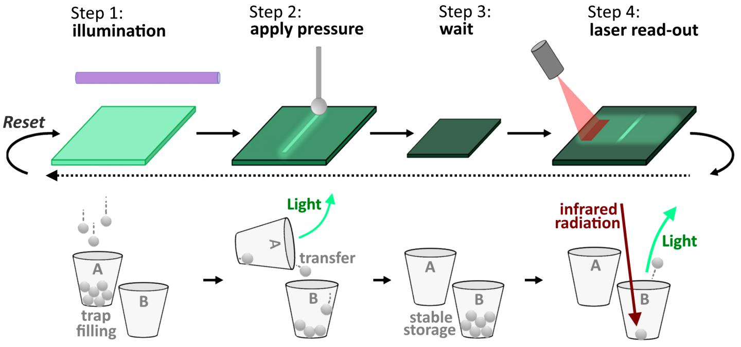
“Mechanoluminescent material was incorporated in polymer plates and briefly exposed to ultraviolet light. During this illumination, defects in the material are populated with electrons originating from the luminescent centers. When pressure is applied to the luminescent material, these electrons are released again from the defects, or traps, as we commonly call them. Some of the released electrons return to the luminescent centers, with the emission of light as a consequence. This is the standard mechanoluminescence process. A fraction of the released electrons are however transferred to other types of defects. Those defects are called ‘deep traps’ as the electrons are not easily released again, except when infrared light is used. By scanning an infrared laser beam over the sample surface, those areas where electrons had been stored after the application of pressure light up as soon as the infrared photons are kicking them out of their traps. The electrons return to the luminescent center and the material locally lights up again.”
The key message is that different types of defects in the luminescent material can have different responses to stimuli like heat, pressure or light. In this way, electrons can be moved by pressure – the “write” action – and then be “read out” by optical means. Because the deep traps are very stable, the researchers were able to demonstrate that the signal was still visible after waiting three days between the “write” and “read” steps. But the researchers’ work is far from finished. First, they want to improve the sensitivity of the mechanoluminescent material and optimize the write and read conditions. Second, they plan to bring the material to specific applications where optimal use of the memory function can be exploited. “Think of components of buildings, vehicles or infrastructure where constant monitoring is not possible”, Smet adds. “Furthermore, this approach will not only help the development of new pressure indicating materials. This work also revealed that the defects responsible in the energy storage and read out process are not of a single type. This insight will prove beneficial for the development of other light-emitting materials, where researchers use defects for other purposes or try even to suppress the occurrence of defects. Think of LED lamps which might be even more efficient in the conversion of electricity to light, brighter glow-in-the-dark materials or faster scintillating materials used in medical imaging,” the scientists forecast.
###
Media Contact
Philippe Smet
[email protected]
Related Journal Article
http://dx.


