X-ray imaging visualizes hidden structures and processes in living cells and organisms. The radiation that consists of highly energy-rich electromagnetic waves, however, has an ionizing effect and may damage the genetic material. This limits the possible observation period. While conventional X-ray images of soft tissue are of low contrast, phase contrast methods produce far better image contrasts at a reduced radiation dose. With higher resolution, however, gentle imaging becomes increasingly difficult, as a higher dose is required. Moreover, the efficiency of the usually applied high-resolution detectors decreases, as a result of which radiation exposure is further increased. So far, high-resolution X-ray phase contrast imaging of living biological specimens has been possible for a few seconds to minutes only, before severe damage is caused by the radiation.
Credit: (Image: Rebecca Spiecker, KIT)
X-ray imaging visualizes hidden structures and processes in living cells and organisms. The radiation that consists of highly energy-rich electromagnetic waves, however, has an ionizing effect and may damage the genetic material. This limits the possible observation period. While conventional X-ray images of soft tissue are of low contrast, phase contrast methods produce far better image contrasts at a reduced radiation dose. With higher resolution, however, gentle imaging becomes increasingly difficult, as a higher dose is required. Moreover, the efficiency of the usually applied high-resolution detectors decreases, as a result of which radiation exposure is further increased. So far, high-resolution X-ray phase contrast imaging of living biological specimens has been possible for a few seconds to minutes only, before severe damage is caused by the radiation.
Researchers from KIT’s Laboratory for Applications of Synchrotron Radiation (LAS), Institute for Photon Science and Synchrotron Radiation, and Physikalisches Institut have now developed a method that uses radiation more efficiently and produces images of micrometer resolution. The method is suited for both living specimens and sensitive materials and opens up new opportunities in biology, biomedicine, and materials sciences. The new system combines X-ray phase contrast with a so-called Bragg magnifier and a photon-counting detector.
Directly Enlarged X-ray Image
“Instead of converting the X-ray image into an image with visible light and enlarging it afterwards, we enlarge it directly,” LAS doctoral researcher Rebecca Spiecker says. “Thanks to this approach, we can use highly efficient large-area detectors.” The researchers use a photon-counting detector with a pixel size of 55 micrometers. Before, the X-ray image of the specimen is enlarged with a so-called Bragg magnifier, as a result of which the resolution of the specimen proper reaches about 1 micrometer. The Bragg magnifier consists of two perfect silicon crystals, whose enlarging effect results from asymmetric diffraction in the silicon crystal lattice. Another big advantage of the Bragg magnifier is the very good optical image transmission. It allows for the nearly loss-free reproduction of all spatial frequencies up to the resolution limit.
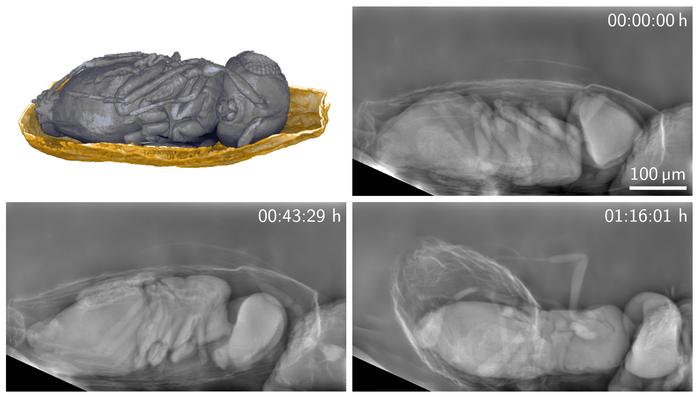
Parasitic Wasps Observed for 30 Minutes
Thanks to the combination of propagation-based X-ray phase contrast with a Bragg magnifier and a photon-counting detector, all of which are optimized for an X-ray energy of 30 kiloelectron-volts (keV), the method reaches about the maximum possible dose efficiency for X-ray phase contrast. This allows for far longer observation times of small living organisms with micrometer resolution. Together with scientists from all over Germany, the researchers demonstrated the method in a pilot study of smallest parasitic wasps. For more than 30 minutes, they observed the wasps in their host eggs and how they emerged from them. “The method is also suited for biomedical applications, an example being the gentle three-dimensional histological investigation of biopsy samples,” Spiecker says. The researchers now plan to further improve the setup, to enlarge the field of view, and to increase mechanical stability for even longer measurements.
Original Publication (Open Access):
Rebecca Spiecker, Pauline Pfeiffer, Adyasha Biswal, Mykola Shcherbinin, Martin Spiecker, Holger Hessdorfer, Mathias Hurst, Yaroslav Zharov, Valerio Bellucci, Tomas Farago, Marcus Zuber, Annette Herz, Angelica Cecilia, Mateusz Czyzycki, Carlos Dias, Dmitri Novikov, Lars Krogmann, Elias Hamann, Thomas van de Kamp, and Tilo Baumbach: Dose-efficient in vivo X-ray phase contrast imaging at micrometer resolution. Optica, 2023. DOI 10.1364/OPTICA.500978
https://doi.org/10.1364/OPTICA.500978
Journal
Optica
DOI
10.1364/OPTICA.500978
Method of Research
Imaging analysis
Subject of Research
Animals
Article Title
Dose-efficient in vivo X-ray phase contrast imaging at micrometer resolution by Bragg magnifiers
Article Publication Date
7-Dec-2023


