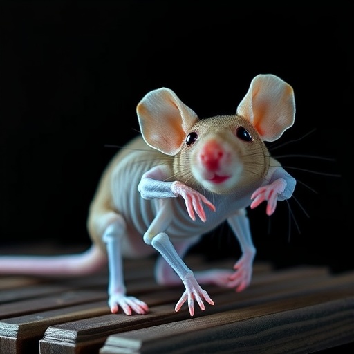
A groundbreaking study has emerged from a collaboration between researchers at the University of Texas Southwestern Medical Center and Harvard Medical School, presenting a novel mouse model that elucidates the complex mechanisms underlying osteogenesis imperfecta (OI), a perplexing genetic bone disorder characterized by brittle bones and frequent fractures. This innovative research focuses on mutations in the Sp7 gene—particularly the arginine-to-cysteine substitution at position 342 (R342C) in mice, which mirrors a similarly pathogenic mutation found at position 316 in humans. By mimicking this mutation in mice, scientists have created an invaluable experimental platform to explore the intricate cellular and molecular dysfunctions driving OI.
Osteogenesis imperfecta has long been associated primarily with defects in the collagen matrix, as mutations impair the biosynthesis of collagen—a vital protein that confers mechanical strength and resilience to bone tissue. However, emerging evidence has implicated the role of osteocytes—the most abundant bone cells that originate from osteoblasts—to be critical in maintaining bone homeostasis. Yet, the extent to which osteocytes contribute to OI pathogenesis remains poorly understood. This novel mouse model offers an unprecedented glimpse into how mutated Sp7 transcription factor affects osteocyte morphology and function.
The Sp7 gene encodes specificity protein 7 (Sp7), a transcription factor fundamental to osteoblast differentiation and bone formation. Mutations in SP7 have been identified in rare subsets of OI patients, exhibiting reduced osteocyte density and morphological abnormalities within bone matrix. These clinical observations informed the precise engineering of the Sp7 R342C mutant mouse using the cutting-edge in vivo genome editing technique known as iGONAD. This allowed researchers to introduce the point mutation at the endogenous locus, ensuring physiological expression and faithful phenotypic modeling.
.adsslot_txs4WeLCDP{ width:728px !important; height:90px !important; }
@media (max-width:1199px) { .adsslot_txs4WeLCDP{ width:468px !important; height:60px !important; } }
@media (max-width:767px) { .adsslot_txs4WeLCDP{ width:320px !important; height:50px !important; } }
ADVERTISEMENT
Subsequent micro-computed tomography (micro-CT) analyses of the femurs of mutant mice revealed compelling skeletal abnormalities. The mutant bones exhibited markedly lower bone mineral density and a reduced fraction of trabecular bone volume—hallmarks of compromised structural integrity. Moreover, cortical porosity in the outer bone layer was significantly elevated, indicating a disruption in cortical bone quality. This phenotype is strikingly congruent with skeletal disorders noted in human patients harboring homozygous Sp7 R316C mutations, underscoring the translational relevance of this model.
Delving deeper, the researchers interrogated the bone remodeling dynamics, a tightly regulated process orchestrated by osteoclast-mediated bone resorption and osteoblast-mediated bone formation. Intriguingly, the Sp7 mutant mice demonstrated an abnormal remodeling balance with increased intracortical remodeling activity. Such dysregulation may contribute to the observed porous cortical bone structure, highlighting a potential uncoupling of osteoblastic and osteoclastic functions in the mutant context.
In tandem, histological assessments revealed a profound reduction in the number of osteocyte dendrites—elongated cellular extensions essential for mechanosensation and intercellular signaling within the bone matrix. These dendrites facilitate the communication network that regulates bone turnover and adaptation. The paucity of osteocyte dendrites in mutant mice suggests impaired mechanotransduction, which could exacerbate bone fragility. Furthermore, elevated apoptosis rates among osteocytes were documented, indicating compromised cell survival pathways that may undermine bone maintenance.
Genomic profiling using ribonucleic acid sequencing (RNA-seq) provided additional mechanistic insights. Comparison of osteocyte-enriched bone cell populations between mutant and wild-type mice revealed widespread transcriptomic alterations. Specifically, over a thousand genes demonstrated increased expression while nearly a thousand were downregulated. Among them, 22 genes critically associated with osteocyte function were disrupted, signifying profound molecular perturbations induced by the Sp7 mutation. Notably, Tnfsf11, a gene encoding RANKL—a pivotal cytokine promoting osteoclastogenesis—was significantly upregulated, potentially explaining the escalated bone resorption phenotype.
To disentangle the bidirectional relationship between osteocyte defects and bone resorption, the investigators employed osteoprotegerin-Fc (OPG-Fc), a decoy receptor that inhibits RANKL-mediated osteoclast activation. Treatment of mutant mice with OPG-Fc successfully diminished cortical porosity by curbing excessive bone resorption. However, osteocyte dendrite abnormalities persisted despite normalized remodeling parameters, suggesting that dendritic deficits are intrinsic to osteocyte pathobiology independent of osteoclast activity. This critical observation sheds light on previously unappreciated osteoclast-independent pathways perpetuating bone fragility in OI.
Collectively, these findings underscore that OI pathology involves more than just defective collagen synthesis. The Sp7 R342C mutation orchestrates a cascade of molecular and cellular anomalies within osteocytes, culminating in impaired bone remodeling, structural fragility, and heightened resorptive activity. The interplay between transcriptional dysregulation, osteocyte morphological defects, and apoptotic pathways unveils novel therapeutic targets that transcend conventional approaches aimed solely at collagen restoration.
This work also exemplifies the power of integrating advanced genetic engineering, high-resolution imaging, transcriptomics, and targeted pharmacological interventions to dissect bone disease mechanisms at unprecedented resolution. By faithfully recapitulating human pathological mutations in a murine system, the study fosters a deeper understanding of OI etiology and opens avenues for precision medicine strategies tailored to osteocyte dysfunction.
Importantly, the persistence of osteocyte dendrite defects despite osteoclast inhibition emphasizes the need to develop therapeutic modalities capable of restoring osteocyte connectivity and survival. Enhancing osteocyte health may prove pivotal in stabilizing bone architecture and reducing fracture risk in patients with SP7 mutations. Such targeted interventions would complement existing treatments that focus predominantly on modulating bone resorption.
As the scientific community continues to unravel the complexities of skeletal diseases, studies like these highlight the critical role of transcription factors like Sp7 in orchestrating bone cell function and integrity. They reaffirm that the bone microenvironment is a dynamic ecosystem where subtle genetic alterations can ripple through multiple cellular compartments, reshaping tissue architecture and function in profound ways.
The unveiling of osteoclast-independent osteocyte defects underscores a paradigm shift in our comprehension of bone disorders, challenging researchers and clinicians alike to rethink therapeutic paradigms. With continued multidisciplinary efforts, this knowledge may translate into innovative treatments that enhance quality of life for individuals with OI and related musculoskeletal diseases.
Subject of Research: Animals
Article Title: Osteoclast-independent osteocyte dendrite defects in mice bearing the osteogenesis imperfecta-causing Sp7 R342C mutation
News Publication Date: 19-Jul-2025
References: DOI: 10.1038/s41413-025-00440-1
Image Credits: Dr. Jialiang S. Wang and Dr. Marc N. Wein from Harvard Medical School, USA
Keywords: Orthopedics, Genetics, Life sciences, Cell biology, Molecular biology, Bone diseases, Musculoskeletal system, Animal models, Bone formation, Biotechnology
Tags: bone homeostasis mechanismsbrittle bone disorder studiescollagen matrix biosynthesis defectsexperimental models for genetic disordersgenetic basis of bone diseasesgenetically engineered mouse modelimplications of osteocyte morphologynovel research collaboration in geneticsosteocyte function in bone healthosteogenesis imperfecta researchSp7 gene mutationstranscription factors in bone development


