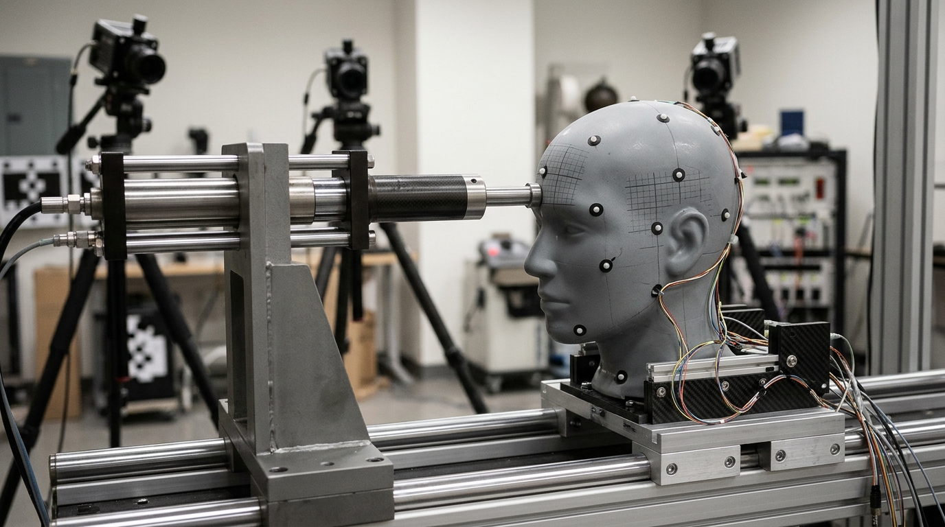
Frontal impacts produce consistently larger peak intracranial displacements and tissue strains than occipital impacts regardless of impactor compliance or gross kinematics, according to dynamic measurements of discrete anatomical regions in post-mortem human subject heads. The findings, reported in the Annals of Biomedical Engineering, demonstrate that internal mechanics within the human skull are sensitive to impact direction, impact site, and local boundary geometry.
Traumatic brain injury frequently arises from falls, sports collisions, violence, and transportation or industrial events, primarily driven by inertial loading that induces intracranial deformations, rotations, tissue strains, and resulting neuropathology. Computational tools such as finite element models are widely utilized to simulate these biomechanical responses and predict injury patterns, yet their accuracy relies directly on biofidelic experimental data capturing in situ brain–skull kinematics and regional deformation, which have historically remained limited.
To establish these experimental targets, the research team, led by Jean-Jacques Demiannay, Oren E. Petel, and colleagues, evaluated the dynamic response of the human brain when subjected to systematically varied head impact severities, directions, and impactor compliances. The investigators employed high-speed X-ray cinematography to record and quantify how discrete anatomical regions moved within post-mortem human subject specimens during each impact condition.
From these high-speed kinematic recordings, the researchers derived time histories of brain–skull relative displacement and tissue strain specifically formatted for finite element model validation and inter-model comparisons. Detailed regional analysis across the specimens showed that intracranial deformation was spatially non-uniform, with the greatest magnitudes of displacement and strain consistently concentrating in superior brain regions.
The investigators noted that the elevated deformations observed superiorly may be related to differences in local skull and meningeal geometry relative to the skull base. By providing experimentally grounded kinematic datasets, the authors stated that these measurements offer validation targets to improve finite element models, while advancing the understanding of neuropathology evolution and aiding the development of head protection strategies tailored to directional and regional vulnerabilities.
Subject of Research: Biomechanical measurement of intracranial brain deformation during head impacts
Article Title: Measurements of Intracranial Brain Deformation in Post-mortem Human Subject Specimens Subjected to Variable Impact Conditions
Article References: Demiannay, J.-J., Brannen, M., Xu, S., Azadi, A. H., Dehghan, P., Goodwin, S., Yahiaoui-Djerboua, W., Taylor, R., Poon, K., Brien, S., Vassilyadi, M., Cusimano, M. D., Karton, C., Hoshizaki, B., & Petel, O. E. (2026). Measurements of Intracranial Brain Deformation in Post-mortem Human Subject Specimens Subjected to Variable Impact Conditions. Annals of Biomedical Engineering. https://doi.org/10.1007/s10439-026-04419-9
Image Credits: AI Generated
DOI: 10.1007/s10439-026-04419-9
Keywords: traumatic brain injury, biomechanics, brain deformation, finite element models, impact kinematics, post-mortem human subjects, high-speed X-ray cinematography, intracranial strain, Measurements, Intracranial, Brain, Deformation
News Source: Cassandra Pierce. (November 7, 2026). Frontal Impacts Cause Greater Brain Deformation in Human Head Specimen Tests. Scienmag.