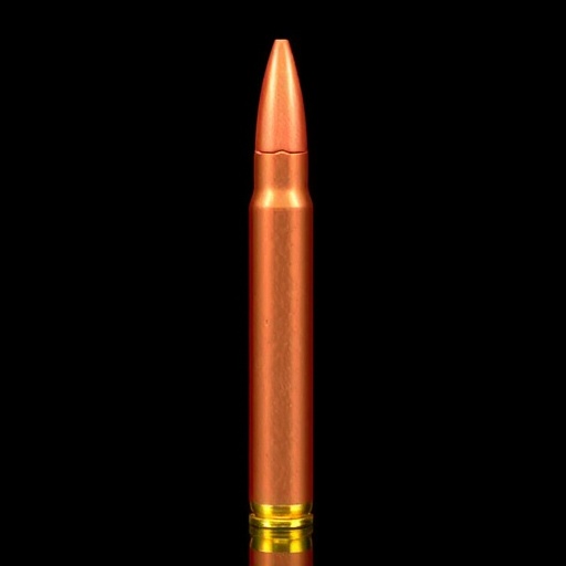

In a groundbreaking advancement poised to revolutionize forensic ballistics, researchers have unveiled an innovative method for analyzing shotgun pellets’ material composition using dual-energy computed tomography (DECT). This technique leverages the nuanced differences in energy absorption of materials to enhance the precision of forensic investigations. Shotgun injuries, frequently encountered in violent crimes and hunting accidents alike, present unique challenges due to the diverse composition of pellets and their complex interactions with surrounding tissues and materials. Understanding these intricacies has now become significantly more feasible thanks to this state-of-the-art imaging technology.
For decades, forensic investigators have grappled with the limitations of traditional imaging methods when it came to identifying the precise material makeup of shotgun pellets embedded within bodies or crime scenes. Conventional computed tomography (CT) scans have provided structural insights but fallen short in differentiating between metals like lead, steel, or other alloys, all commonly used in shotgun ammunitions. DECT’s innovative approach, employing two distinct X-ray energy spectra, heightens contrast and enables material-specific identification, thereby offering a powerful tool in forensic diagnostics.
The research team demonstrated that DECT could non-invasively distinguish between various pellet materials by analyzing their attenuation properties at different energy levels. This capability profoundly impacts forensic reconstructions, enabling experts to determine the exact type of ammunition used without subjecting the specimen to destructive testing. As law enforcement agencies worldwide seek ever more reliable and scientifically rigorous methods to support criminal investigations, this technology is poised to become indispensable in forensic medicine.
.adsslot_ehwaOSlZmE{width:728px !important;height:90px !important;}
@media(max-width:1199px){ .adsslot_ehwaOSlZmE{width:468px !important;height:60px !important;}
}
@media(max-width:767px){ .adsslot_ehwaOSlZmE{width:320px !important;height:50px !important;}
}
ADVERTISEMENT
What makes DECT especially suited for shotgun pellet examination is its ability to generate what is termed as “material-specific images.” These images exploit the differing x-ray absorption characteristics of materials at high and low energy settings. By applying sophisticated algorithms to these data, the resulting images transcend mere shape and density, revealing unique “signatures” indicative of the pellet’s elemental composition. This enhancement allows forensic experts to not only pinpoint pellet location within the body but also discern between toxic lead projectiles and inert steel variants, which has far-reaching implications for medical treatment and legal proceedings.
This novel application of DECT extends its utility beyond forensic analysis into the medico-legal evaluation of gunshot wounds. The accurate characterization of pellet materials facilitates tailored clinical interventions, as different metals provoke varying biological responses when lodged in human tissue. For instance, lead toxicity is a well-documented risk in gunshot injuries involving lead-based ammunition, while steel pellets might present alternative complications. Early and precise material identification aids clinicians in developing appropriate treatment plans, potentially reducing patient morbidity and enhancing recovery outcomes in trauma care.
Moreover, the non-destructive nature of DECT represents a leap forward in preserving forensic evidence integrity. Traditional methods often require extraction and physical testing of pellets, which risks contamination or alteration. DECT scanning allows forensic pathologists and crime scene investigators to conduct thorough material analyses while maintaining the original state of evidence, a vital consideration in courtroom settings. The enhanced imaging can also assist in ballistic trajectory reconstruction and corroborate witness statements or suspect confessions, bolstering the evidentiary value of forensic findings.
Another facet of this research highlights its contribution to understanding the fabrication and usage trends of shotgun ammunition. By compiling data on pellet material characteristics across different cases, forensic scientists can build comprehensive databases that may link specific manufacturers or ammunition batches to crime scenes. This capability transforms DECT into not just a diagnostic instrument but a strategic intelligence tool, opening new avenues in criminal profiling and forensic investigation techniques.
The research also underscores the technological challenges and limitations associated with employing DECT in forensic contexts. High-resolution dual-energy scanners are sophisticated and require specialized interpretation by trained radiologists and forensic experts. Additionally, overlapping attenuation values for some alloys might confound the identification process, necessitating continual refinement of imaging protocols and analytical algorithms. Nevertheless, the potential benefits greatly outweigh these obstacles, positioning DECT as an emergent standard in forensic radiology.
In terms of practical implementation, the study advocates for equipping forensic laboratories and medical examiner offices with DECT capabilities. The initial investment in equipment and training is balanced by the enhanced accuracy, safety, and efficiency that this technology brings to pellet analysis. Integrating DECT with existing forensic workflows would enable seamless adoption, ensuring timely and conclusive results that significantly aid both criminal justice and medical communities.
Crucially, the authors emphasize the interdisciplinary nature of this advancement. Radiologists, forensic pathologists, materials scientists, and legal experts must collaborate to harness the full potential of DECT for shotgun pellet examination. Such cooperation fosters comprehensive methodologies that consider technological nuances, biological implications, and evidentiary standards. This holistic approach aligns with modern trends in forensic science, where technological innovation and cross-disciplinary integration drive breakthroughs.
The implications of this research reverberate even further, suggesting that dual-energy computed tomography could be adapted for other forensic applications. Bullet fragments from firearms, shrapnel from explosive devices, and foreign objects in trauma cases may all benefit from similar material-specific imaging. The conceptual framework developed for shotgun pellet analysis thus lays the foundation for a suite of forensic imaging modalities that improve accuracy and objectivity in evidence interpretation.
Additionally, the forensic community anticipates that enhanced material detection will lead to improved legal outcomes. The ability to definitively identify ammunition types could influence case proceedings by strengthening evidence admissibility and reducing ambiguities that defense or prosecution parties might exploit. This scientific precision bolsters confidence in expert testimonies, contributing to fairer and more transparent judicial processes. DECT’s role in achieving these goals marks a pivotal step forward in forensic jurisprudence and criminal investigation.
This transformative technology’s social impact is also noteworthy. By accelerating accurate injury assessment and ammunitions identification, medical and legal systems can respond more swiftly and justly to gun-related incidents. This has potential downstream effects on public health policies, firearm regulations, and community safety initiatives. The fusion of medical imaging innovation with forensic science exemplifies how technology can serve society’s broader interests through enhanced justice and improved trauma care.
In summary, the pioneering use of dual-energy computed tomography to examine forensic shotgun pellets heralds a new era in ballistic forensics. The ability to non-invasively identify pellet materials with high fidelity improves evidence quality, supports clinical treatment, and sharpens investigative accuracy. While the technology demands specialized expertise and resources, its numerous advantages point to widespread adoption in the near future. As researchers continue to refine DECT applications and expand its forensic toolkit, this breakthrough stands as a testament to the power of technological ingenuity in elevating scientific standards within the justice system.
Subject of Research: Forensic examination and material characterization of shotgun pellets using dual-energy computed tomography (DECT).
Article Title: Forensic shotgun pellet examination – material detection with dual-energy computed tomography.
Article References:
Brix, M., Junno, J.A., Lamentausta, E. et al. Forensic shotgun pellet examination – material detection with dual-energy computed tomography. International Journal of Legal Medicine (2025). https://doi.org/10.1007/s00414-025-03553-8
Image Credits: AI Generated
DOI: https://doi.org/10.1007/s00414-025-03553-8
Keywords: Forensic ballistics, dual-energy computed tomography, shotgun pellets, material identification, forensic radiology, gunshot wound analysis, non-destructive testing, forensic imaging
Tags: advanced imaging techniques in forensicsattenuation properties in imagingcrime scene investigation technologiesdifferentiating metals in ammunitiondual-energy computed tomography applicationsforensic ballistics analysisforensic diagnostic advancementsinnovative methods in forensic sciencematerial composition analysis in forensicsnon-invasive forensic investigationsshotgun injuries forensic analysisshotgun pellet material identification

