In a groundbreaking study, researchers have illuminated the complex dynamics of false lumen hemodynamics in Type B aortic dissection, an area that has remained shrouded in ambiguity until now. The work, utilizing advanced Particle Image Velocimetry (PIV) and patient-specific flexible phantoms, provides critical insights into how blood flows within the aorta under pathological conditions. This sophisticated methodology allows the team to not only visualize but also quantify the blood flow patterns that emerge in the false lumen—an area often associated with serious complications such as poor perfusion and potential organ ischemia.
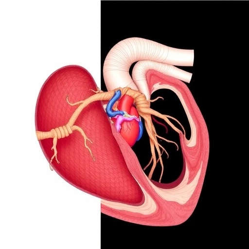
Type B aortic dissection, characterized by a tear in the aorta’s middle layer, leads to the formation of a false lumen where blood can flow unpredictably. The study conducted by Koulogiannis and his associates marks a significant advancement in the understanding of this condition. Historically, the implications of such hemodynamics were not thoroughly understood, primarily due to the complex relationship between flow dynamics and the geometry of the dissection. This research bridges that gap by employing patient-specific models that reflect the true anatomical diversity observed in the human population.
One of the standout features of this research is the use of PIV, a technique that captures the flow of liquid in detail. PIV operates by illuminating particles suspended in the fluid with a laser and capturing images of the flow field. By analyzing the movement of these particles, researchers can extract valuable data about the velocity and direction of blood within the false lumen. This level of detail presents a more nuanced understanding of how different mechanical forces come into play, offering vital information that could impact clinical outcomes.
The use of patient-specific flexible phantoms is another pivotal element of the study, enhancing its realism and applicability in clinical settings. These phantoms are designed to replicate the individual anatomical features of patients who have experienced Type B aortic dissection. By using a model that accurately reflects a patient’s unique circumstances, the researchers can draw more relevant conclusions regarding treatment approaches and potential interventions. This level of customization in models signifies a leap towards personalized medicine, tailoring approaches based on the specifics of each case.
Through their experiments, the research team meticulously documented the complex interactions between blood flow and the structural changes caused by dissection. Their findings reveal that the flow patterns in the false lumen can vary significantly depending on the dissection’s characteristics, such as its size and the extent of the tears. By correlating these findings with data from patient cases, the researchers are laying the groundwork for targeted therapeutic strategies that need to account for these variations.
Moreover, this research opens up avenues for predicting complications associated with Type B aortic dissection. Understanding the hemodynamics within the false lumen could lead to earlier interventions and improve surgical outcomes. For clinicians, these insights can prove invaluable; the ability to anticipate adverse events could change the management framework surrounding this potentially life-threatening condition.
The project not only enhances understanding from a theoretical standpoint but also has tangible implications for future research endeavors. By establishing a reliable method to simulate and analyze false lumen hemodynamics, subsequent studies can build on this work, potentially exploring further variables such as varying blood pressures, varied heart rates, and the introduction of external forces. Each of these factors would contribute crucial knowledge that could help clinicians develop a comprehensive understanding of Type B dissections.
As a result, this research stands to make significant contributions to both basic science and clinical practice. With the potential to refine treatment protocols, it may also pave the way for developing novel interventions or surgical techniques that mitigate the heightened risks presented by the false lumen. Additionally, the visibility given to fluid dynamics in this context could lead to enhanced diagnostic capabilities, helping healthcare professionals identify high-risk patients more effectively.
The investigative team, composed of notable figures in the field, emphasizes the importance of integrating advanced technology and clinical insight. Their collaborative approach underscores the necessity of merging engineering techniques like PIV with clinical expertise to unravel the complexities of conditions such as aortic dissection. This partnership between disciplines highlights the trajectory towards more innovative and effective healthcare solutions that arise from interdisciplinary collaboration.
The implications of this work extend beyond aortic dissection, potentially inspiring similar methodologies across various cardiovascular research topics. By showcasing the potential of high-fidelity models and real-time fluid dynamics visualization, it invites a reconsideration of how other diseases might be studied and understood. The hope is that this study will not only shed light on aortic dissections but encourage a broader conversation about the importance of detailed hemodynamic analyses in medicine.
In conclusion, the pioneering research conducted by Koulogiannis and colleagues signals a new frontier in the understanding of Type B aortic dissection. Their innovative techniques and comprehensive analysis lay a foundation for future studies and clinical practices aimed at reducing the morbidity associated with this condition. As the fields of engineering and medicine continue to intertwine, the upcoming generation of health professionals are better equipped to tackle the challenges posed by complex cardiovascular diseases.
With the publication of these findings in Annals of Biomedical Engineering, the scientific community is now poised to engage with new data that could fundamentally transform how Type B aortic dissection is approached, treated, and understood by both clinicians and patients alike.
Subject of Research: Aortic dissection hemodynamics using advanced imaging techniques.
Article Title: False Lumen Haemodynamics in Type B Aortic Dissection: An in Vitro Study Using PIV and Patient-Specific Flexible Phantoms.
Article References:
Koulogiannis, A., Li, Q., Homer-Vanniasinkam, S. et al. False Lumen Haemodynamics in Type B Aortic Dissection: An in Vitro Study Using PIV and Patient-Specific Flexible Phantoms.
Ann Biomed Eng (2025). https://doi.org/10.1007/s10439-025-03875-z
Image Credits: AI Generated
DOI: https://doi.org/10.1007/s10439-025-03875-z
Keywords: Aortic dissection, hemodynamics, Particle Image Velocimetry, patient-specific phantoms, cardiovascular research.
Tags: advanced imaging techniques in vascular researchanatomical diversity in dissection studiesaortic dissection research advancementsblood flow quantification methodscomplications of aortic dissectionfalse lumen blood flow patternsfluid dynamics in aortic dissectionorgan ischemia in aortic conditionsParticle Image Velocimetry applicationpatient-specific aortic modelsType B aortic dissection hemodynamicsunderstanding aortic tear implications
