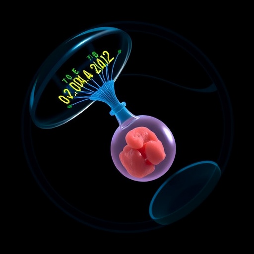
In a groundbreaking achievement, researchers at the Institute for Bioengineering of Catalonia (IBEC), working in close collaboration with the Dexeus University Hospital, have captured the implantation of a human embryo in real time and in three dimensions for the very first time. This pioneering study offers an unprecedented view into one of the most critical and intricate stages of human reproduction—the moment when an embryo embeds itself into the uterine lining, initiating pregnancy. Until now, the intricate mechanics and forces underlying this process in humans have remained elusive, primarily understood through static images taken at isolated phases rather than continuous observation.
Implantation failure is a major contributing factor to infertility worldwide, accounting for approximately 60% of spontaneous miscarriages. By shedding light on the physical and biochemical interactions during implantation, this discovery promises to revolutionize assisted reproductive technologies and improve fertility outcomes. The revelations from this work not only clarify fundamental biological mechanisms but also open new avenues for addressing reproductive disorders by focusing on the embryo’s mechanical dialogue with the maternal environment.
The study reveals that human embryos exert significant mechanical forces as they invade the uterine tissue. Samuel Ojosnegros, principal investigator at IBEC and lead author of the research, describes the process as surprisingly invasive. He explains that embryos do not passively attach but actively burrow into the uterus, leveraging considerable traction forces to integrate fully with the maternal tissue. This invasive behavior corroborates clinical observations where many women experience abdominal discomfort and slight bleeding during implantation, phenomena now better understood through direct visualization.
.adsslot_x0zHcNliaZ{width:728px !important;height:90px !important;}
@media(max-width:1199px){ .adsslot_x0zHcNliaZ{width:468px !important;height:60px !important;}
}
@media(max-width:767px){ .adsslot_x0zHcNliaZ{width:320px !important;height:50px !important;}
}
ADVERTISEMENT
Essential to this invasive process is the combination of biochemical and mechanical factors. The embryo secretes proteolytic enzymes that degrade the surrounding extracellular matrix, facilitating tissue breakdown. Concurrently, the embryo applies physical forces to remodel and penetrate the dense, collagen-rich fibrous tissue of the uterine lining. Collagen, known for its structural rigidity in tendons and cartilage, poses a formidable barrier that the embryo must navigate. By mechanically manipulating this matrix, the embryo creates a path that not only allows its embedding but also promotes the formation of specialized tissues that will connect to the maternal blood supply, ensuring nutrition and growth.
Crucially, the research identifies that the embryo’s traction forces lead to substantial remodeling of the uterine matrix. This mechanical interaction is bidirectional; the embryo not only exerts force but also responds to external mechanical cues present in its environment. Co-first author Amélie Godeau highlights the embryonic sensitivity to the mechanical milieu, suggesting that uterine contractions experienced naturally in vivo could influence the success and pattern of implantation. Such mechanosensitivity underscores the importance of physical signals alongside chemical interactions, broadening the conventional understanding of embryo–uterus communication.
To dissect these complex dynamics, the IBEC team engineered a novel in vitro platform that replicates the uterine environment outside the human body with high fidelity. This platform consists of a bioengineered gel matrix mimicking the collagenous composition of uterine tissue, supplemented with proteins essential for embryonic development. By employing advanced real-time fluorescence imaging techniques, the researchers quantified the mechanical forces and spatial displacements involved as the embryo migrated, embedded, and expanded within this controlled setting. This system bridges the gap between in vivo complexity and in vitro controllability, enabling detailed mechanistic studies impossible before.
Comparative experiments involving both human and murine embryos uncovered species-specific implantation behaviors. In the mouse model, the embryo adheres to the uterine surface and the uterus responds by folding around it, encasing the embryo within a specialized uterine crypt. In stark contrast, human embryos penetrate deeply, infiltrating the uterine lining to begin radial growth from the interior outward. These differences illuminate evolutionary variations in reproductive strategies and have profound implications for interpreting animal models of human implantation.
Beyond fundamental science, this research holds promise for clinical translation. By elucidating the physical underpinnings of implantation failure, which remains a stubborn barrier in infertility treatment, novel interventions could be designed to support or mimic the mechanical environment conducive to embryo embedding. This might include refining culture systems in assisted reproductive technologies or developing therapies targeting the extracellular matrix to optimize uterine receptivity.
The multi-institutional collaboration integrated expertise from the Biomimetic Systems for Cell Engineering group at IBEC, the Barcelona Stem Cell Bank, University of Barcelona, Tel Aviv University, the Biomedical Research Networking Centre (CIBER), and IRB Barcelona. Tissue samples and embryos used in the study were ethically donated by the Dexeus University Hospital, ensuring adherence to strict research regulations and fostering scientific rigor.
The insights from this work challenge and enrich longstanding paradigms about embryo implantation, emphasizing its active mechanical nature. By demonstrating that embryos not only chemically prepare but physically sculpt their implantation niche, the study propels forward a new interdisciplinary field straddling bioengineering, developmental biology, and reproductive medicine. It promises to inspire future research aiming to unravel the finely tuned orchestration enabling new human life from its earliest moments.
Subject of Research: Human embryos
Article Title: Traction force and mechanosensitivity mediate species-specific implantation patterns in human and mouse embryos
News Publication Date: 15-Aug-2025
Image Credits: Institute for Bioengineering of Catalonia (IBEC)
Keywords: Human reproduction, Embryo implantation
Tags: assisted reproductive technologiesbioengineering in reproductive healthclinical implications of embryo researchembryo mechanical forceshuman embryo implantationinfertility research breakthroughsmaternal-embryo interactionmechanisms of implantationmiscarriage causes and solutionsreal-time embryo observationreproductive technology advancementsthree-dimensional imaging of embryos


