Molecular ‘fingerprint’ for tissue taken from first isotope-enriched mouse has huge potential for scientific breakthroughs, as well as improved medical implants. Earliest research based on data has already revealed that a molecule thought to exist for repairing DNA may also in fact trigger bone formation.
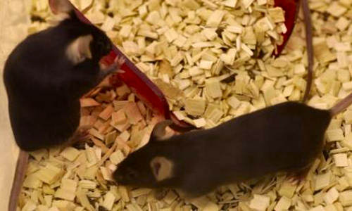
Scientists have created a ‘heavy’ mouse, the world’s first animal enriched with heavy but non-radioactive isotopes – enabling them to capture in unprecedented detail the molecular structure of natural tissue by reading the magnetism inherent in the isotopes.
This data has been used to grow biological tissue in the lab practically identical to native tissue, which can be manipulated and analysed in ways impossible with natural samples. Researchers say the approach has huge potential for scientific and medical breakthroughs: lab-grown tissue could be used to replace heart valves, for example.
In fact, with their earliest research on the new in vitro tissue, the team have discovered that poly(ADP ribose) (PAR) – a molecule believed to only exist inside a cell for the purpose of repairing DNA – not only travels outside cells but may trigger bone mineralisation.
“It was crazy to see PAR behaving in this way; it took six months of detailed analysis and many more experiments to convince ourselves,” said Dr Melinda Duer from Cambridge’s Department of Chemistry, who led the study, published today in the journal Science.
“I think this is just the first of many discoveries that will stem from the heavy mouse. Isotope-enriched proteins and cells are fairly commonplace now, but the leap to a whole animal is a big one.
“The heavier nuclei in the carbon isotopes changes the rate of chemical reactions, and many people – myself initially included – didn’t believe you could enrich a whole animal with them. But it worked beautifully,” she said.
The research, funded by the Biotechnology and Biological Sciences Research Council and British Heart Foundation, could lead to improved success rates for medical implants and reduce the need for animals in research, as well as opening up an entirely new approach for biochemical investigation.
The team used a technique called Nuclear Magnetic Resonance spectroscopy (NMR) that can read the magnetic nuclei found in certain isotopes, such as carbon-13 – which has one neutron more than most carbon.
But carbon-13 makes up only 1% of the carbon in our bodies, nowhere near enough to do useful NMR. However, the researchers managed to get the carbon of a mouse up to 20% carbon-13.
So how do you make a heavy mouse? Perhaps obviously, you feed it a lot. “We used mouse feed rich in carbon-13 and let the mouse eat as much as it liked,” said Duer. “It sounds strange, but no one had thought to do it before. Maybe everyone had assumed it wouldn’t work, I certainly got some odd looks from colleagues.”
Using NMR analysis of the mouse tissue to map the distance between the carbon atoms and reveal atomic structures, researchers were able to create a ‘gold standard’ reference for growing tissue in the lab – a fingerprint of the atomic networks that are the basis of proteins in our biology.
The team shared this with scientists Rakesh Rajan and Dr Roger Brooks in the Division of Trauma and Orthopaedic Surgery at the University’s Department of Surgery, who used the Duer group’s NMR ‘spectra’ maps to refine cell cultures and produce exceptional lab-grown tissue that looks near identical to real tissue.
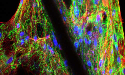
When comparing microscopy images of the new lab-grown tissue with native tissue, Duer says she has yet to find a biologist who can tell the difference. “We found that once you get it right at a molecular level, the rest looks after itself,” she said.
The new techniques allow scientists to go beyond the nanoscopic, which has been the limit for tissue analysis, and into the atomic. “We could see signals in the NMR data for our lab-grown tissue, extra intensities that – when matched with the heavy mouse data – revealed where proteins hadn’t folded up properly,” said Brooks.
This kind of ‘misfolding’ is almost impossible to detect through microscopes, but could result in host rejection if the tissue were to be implanted. “Through a process of repeat NMR comparisons we were able to modify the lab tissue until it looked near identical with NMR and under the microscope,” explained Brooks.
Not only have the researchers developed these techniques, they also managed to strike scientific gold during some of their first experiments on lab-grown tissue matched to the heavy mouse.
Using bone-forming cells, the group grew collagen tissue in the lab to look at how bone is developed. While using the new method to look for sugars, they found signs of molecules that shouldn’t be there – the closest thing these molecules resembled was DNA.
After further research, the team were forced to reach a very surprising conclusion: the molecule was poly(ADP ribose), or PAR, previously only thought to be found inside cells where its purpose in life is flagging damaged DNA for repair.
“Not only is PAR there, and leaving the cells entirely, but once it’s in the surrounding matrix it’s perfectly designed to start pulling together the calcium and phosphate that make up bone crystals,” explained Duer.
This is happening at the exact same time the cells start laying down the organic matrix to house the mineral crystals that form bone, says Duer. In the intervening six months, a staining test for PAR had been developed, so the team checked again on bone growth taken straight from the animal.
“When the results came back, even I couldn’t believe it! The bone tissue was stained everywhere,” said Duer.
The team had discovered that the molecule listed in all the textbooks as the deft surgeon of DNA also moonlights as an engine of bone production.
“The fact that we are already making such remarkable discoveries using the techniques that have been developed as a result of the heavy mouse is hugely exciting, and shows the enormous potential of this approach,” said Duer.
“We’re now looking at blood vessels to see if lab-grown tissue could be used for replacement arteries and heart valves – and to see if we can find the molecules that trigger calcification of the arteries, as well as calcification of bone.”
“One mouse on a specific diet might end up rewriting the textbooks.”
Story Source:
The above story is based on materials provided by University of Cambridge.


