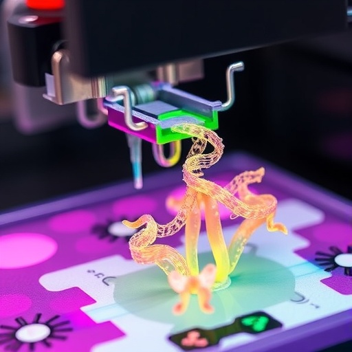
In a groundbreaking advance that promises to reshape the landscape of bioprinting, researchers have unveiled a novel methodology for rapid volumetric bioprinting (VBP) of pristine protein-based bioinks. This innovative approach leverages the inherent tyrosine groups in natural proteins such as silk, gelatin, and decellularized extracellular matrix (dECM) to generate complex, three-dimensional biological constructs swiftly and with high fidelity. Unlike traditional layer-by-layer bioprinting techniques, volumetric bioprinting modifies spatial illumination patterns within a pre-defined build volume, resulting in near-instantaneous photopolymerization that can revolutionize tissue engineering and regenerative medicine.
Conventional VBP systems, while remarkably fast, have struggled with the limited palette of suitable bioinks, primarily relying on chemically modified polymers that often lose native bioactivity or require post-processing steps to restore functionality. The recent work spearheaded by Xie and colleagues introduces an innovative photoinitiator system based on tris(2,2-bipyridyl)dichlororuthenium(II) hexahydrate paired with sodium persulfate, which facilitates the rapid crosslinking of unmodified protein-based bioinks without compromising their pristine state. This critical advancement opens pathways for fabricating biomimetic 3D architectures compatible with further biological or chemical modifications, thereby expanding the utility of volumetric bioprinting in clinical and biomedical settings.
At the heart of this approach lies the exploitation of tyrosine residues naturally abundant in proteins such as silk fibroin, silk sericin, and gelatin. The ruthenium-based photoinitiator system catalyzes a rapid oxidative coupling of these tyrosine groups upon exposure to visible light within seconds, enabling the formation of robust hydrogel networks without introducing foreign chemical groups or extensive preprocessing. This retains the intrinsic biological cues and mechanical properties vital for cell adhesion, proliferation, and differentiation, representing a paradigm shift in biofabrication where biological relevance meets manufacturing efficiency.
The volumetrically printed constructs derived from silk sericin exhibit remarkable shape-memory effects, showcasing reversible shrinkage and expansion behaviors. This property is particularly intriguing for applications requiring dynamic adaptability such as soft robotics, implantable devices, or responsive tissue scaffolds. By contrast, silk fibroin-based bioinks produce constructs with a broad range of tunable mechanical properties, spanning from a few hundred pascals to hundreds of megapascals, thus enabling customization to match the mechanical milieu of various native tissues including cartilage, tendon, or bone.
Perhaps most compelling is the cytocompatibility demonstrated by these protein-based hydrogels. Cellular viability and proliferation assays confirm that both silk- and dECM-based volumetrically printed constructs support living cells without eliciting toxic responses or impairing cellular functions. This compatibility is crucial for future applications in tissue engineering and regenerative therapies, where bioinks must support not just structural roles but also active cellular processes.
The significance of these advances is underscored by the detailed protocols provided by the research team, which include comprehensive steps for bioink preparation, volumetric illumination pattern design, photopolymerization, and post-processing characterization. This standardized methodology ensures reproducibility and scalability, critical for translational research and industrial adoption. The ability to rapidly produce intricate 3D shapes with high resolution facilitates the creation of anatomically matched implants or organotypic models for drug screening and disease modeling.
In terms of clinical applicability, volumetrically printed dECM constructs hold promise for personalized medicine due to their origin from native tissue matrices, offering an environment highly conducive to cell signaling and regeneration. The integration of VBP with these bioinks circumvents the limitations of slow, laborious bioprinting processes, accelerating the journey from design to functional tissue constructs ready for implantation or laboratory study.
Moreover, the new photoinitiator chemistry employed is notable for its visible light activation, reducing potential damage to encapsulated cells compared to ultraviolet-based systems frequently used in photopolymerization. This enhances cell survival rates during printing, an essential factor for creating viable tissue analogs that can thrive post-fabrication.
The robustness of the methodology is further demonstrated through various validation experiments assessing mechanical strength, swelling behavior, and long-term stability of printed constructs. These characterizations affirm the capability of volumetrically printed structures to withstand physiological stresses and maintain functional integrity over time, factors crucial for their deployment in living organisms.
Beyond biomedical uses, such versatile volumetrically bioprinted protein-based hydrogels exhibit potential in soft robotics and bioelectronics. Their biocompatible and tunable properties could be harnessed to design biohybrid devices that seamlessly interface with human tissue or environmental stimuli, pushing the frontiers of bio-integrated technology.
Notably, the new VBP platform holds the potential to democratize bioprinting by simplifying the hardware and material requirements. The use of unmodified, readily available natural bioinks reduces costs and complexity, while the rapid printing times enhance throughput. This positions VBP as a disruptive technology capable of addressing urgent needs in personalized healthcare and pharmaceutical research.
In conclusion, the work by Xie, Lian, Zhang, and collaborators heralds a new era in bioprinting where speed, biological fidelity, and versatility converge. The ability to volumetrically print pristine protein-based bioinks without chemical modification unlocks unprecedented opportunities for fabricating living tissues and functional architectures with clinical relevance. As this technology matures, it may redefine approaches to tissue engineering, accelerate drug development pipelines, and inspire innovations across biomedical engineering disciplines.
This breakthrough not only surmounts longstanding barriers in biofabrication but also aligns with broader efforts to harness natural biomaterials in regenerative medicine. By marrying the chemistry of ruthenium-catalyzed photopolymerization with the intricate biology of native proteins, the researchers offer a blueprint for creating biomimetic constructs that can emulate the dynamic complexity of living systems with unparalleled speed and precision. The future of biofabrication may well hinge on such transformative methodologies, bridging the divide between engineered constructs and living tissue analogs.
Subject of Research:
Rapid volumetric bioprinting of unmodified, pristine protein-based bioinks using a novel photoinitiator system.
Article Title:
Rapid volumetric bioprinting of pristine protein-based (bio)inks.
Article References:
Xie, M., Lian, L., Zhang, Z. et al. Rapid volumetric bioprinting of pristine protein-based (bio)inks. Nat Protoc (2026). https://doi.org/10.1038/s41596-026-01341-1
Image Credits:
AI Generated
DOI:
https://doi.org/10.1038/s41596-026-01341-1
Tags: 2-bipyridyl)dichlororuthenium(II) hexahydrate photoinitiatorbiomimetic tissue engineering scaffoldsfast volumetric bioprinting of protein inkspristine protein-based bioinksrapid 3D bioprinting techniquessodium persulfate crosslinking systemtris(2tyrosine-mediated photopolymerizationunmodified natural protein bioinksvolumetric bioprinting for regenerative medicine
