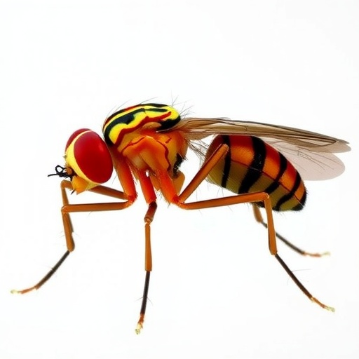
Recent breakthroughs in the study of Drosophila motion vision have illuminated long-standing puzzles about how flies interpret complex visual information. By meticulously investigating the spatial integration patterns of directionally selective T4 neurons, scientists have bridged crucial gaps that unite local circuit mechanisms with the broader optic flow processing observed in larger flies, revealing a fundamental principle that governs visual computation.
The research at hand uncovers a universal sampling rule for T4 neurons, a discovery that profoundly clarifies how the hexagonal arrangement of photoreceptors on the fly’s compound eye underpins the directional tuning of neurons in the lobula plate, a key motion processing center. This sampling rule ensures that each T4 neuron integrates signals from a precise hexagonal unit of medulla columns, adhering closely to the eye’s innate coordinate system. This finding effectively reconciles various behavioral and physiological observations previously noted across different fly species.
For years, studies had produced seemingly disparate results about how local preferred directions (PDs) of motion-sensitive neurons align with the intricate layout of the fly’s eye. Behavioral experiments demonstrated that maximal responses were oriented along rows of ommatidia, while recordings from lobula plate tangential cells in larger flies showed PDs reflecting the skewed geometry of the hexagonal lattice. Now, these phenomena are explained through a common anatomical framework where dendritic orientation and eye structure shape the directional selectivity of T4 neurons, elaborating a cohesive narrative that connects the microcircuitry to global optic flow sensing.
.adsslot_xWa2duBnDj{width:728px !important;height:90px !important;}
@media(max-width:1199px){ .adsslot_xWa2duBnDj{width:468px !important;height:60px !important;}
}
@media(max-width:767px){ .adsslot_xWa2duBnDj{width:320px !important;height:50px !important;}
}
ADVERTISEMENT
Moreover, the directional tuning extends beyond cardinal axes to incorporate nuanced diagonal sensitivities particularly in dorsal and frontal visual fields, as highlighted by studies of looming-sensitive neurons and interneurons with adapting PDs. This aligns with evidence from electrophysiological recordings of HS (Horizontal System) and VS (Vertical System) neurons, further supporting the universality of the underlying sampling mechanism and its predictive power in identifying non-canonical translation axes in optic flow processing.
Complementing these insights, the neuronal responses recorded from T4 and T5 axonal layers show smoothly varying direction preferences across lobula plate strata, forming spatial patterns that resemble translation-like optic flow fields. This layered organization consolidates the notion that local dendritic structure, guided by anatomical constraints, scales up to support global computations essential for flight navigation and motion perception in flies.
Importantly, the functional similarities between T4 and T5—ON and OFF motion detecting counterparts—emerge from shared developmental rules that organize connectivity in the medulla and lobula neuropils, respectively. Despite the structural and functional complexity, RNA sequencing data reveal that all eight T4 and T5 subtypes possess remarkably similar transcriptional profiles, indicating that subtle differences in dendritic orientation rather than fundamental genetic heterogeneity likely drive their distinct roles.
The research also emphasizes the vital role of asymmetrical dendritic wiring in establishing directional selectivity, with each presynaptic neuron targeting specific dendritic locations on T4 and T5 neurons. However, the developmental mechanisms guiding this intricate wiring remain elusive. The discovery of a universal hexagonal sampling motif points toward a conserved developmental program that acts in concert with processes determining dendritic orientation to shape the emergent computational architecture.
Beyond Drosophila, the implications of this work resonate across arthropods, many of which share conserved optic lobe features and motion-sensitive neuron types. Given the extraordinary diversity of compound eye structures across these species, the study provokes intriguing questions about how eye anatomy might universally dictate motion detection strategies and behavioral responses, suggesting that evolutionary pressures sculpt sensory processing in tandem with morphology.
Overall, this research unites a broad spectrum of physiological, anatomical, and computational findings into a cohesive framework that elucidates how the structure of the fly’s eye directly shapes the functional properties of neurons responsible for motion detection. By integrating insights across scales—from the hexagonal lattice of eye facets to large-scale optic flow fields—this work advances our understanding of sensory computation and sets the stage for exploring motion vision in other species with complex compound eyes.
These findings highlight the interdisciplinary nature of contemporary neuroscience, where detailed anatomical reconstructions, electrophysiological recordings, and developmental gene expression analyses converge to unlock the principles underlying perception. The study’s comprehensive approach offers a powerful model system for dissecting how local circuits interplay with global sensory representations, fundamental to navigating dynamic environments.
In sum, the elucidation of this universal sampling rule in T4 neurons bridges years of disparate observations in the field and provides an elegant mechanistic explanation for how directional selectivity emerges from the intricate dance between eye structure and neuronal wiring. This advance paves the way for novel explorations into how sensory systems are tailored by evolution to suit their ecological niches, influencing behavior and survival.
As neuroscientists continue to unravel the complexities of visual processing, the revelations stemming from Drosophila serve as a testament to the power of model organisms in shedding light on fundamental principles with broad biological significance. The interplay between anatomy and function unveiled here marks a milestone in our journey toward comprehending the neural basis of motion perception.
Beyond the immediate implications for fly vision, these insights inspire further inquiry into how universal principles of spatial sampling and circuit organization manifest in other sensory modalities and animal taxa. Understanding these principles could ultimately deepen our grasp of neural computation across the animal kingdom.
Subject of Research:
The relationship between the compound eye structure and the directional selectivity of motion-sensitive neurons (T4 and T5) in Drosophila, elucidating mechanisms underlying local and global optic flow processing.
Article Title:
Eye structure shapes neuron function in Drosophila motion vision.
Article References:
Zhao, A., Gruntman, E., Nern, A. et al. Eye structure shapes neuron function in Drosophila motion vision. Nature (2025). https://doi.org/10.1038/s41586-025-09276-5
Image Credits: AI Generated
Tags: behavioral experiments on fly visioncompound eye structure and functiondirectional tuning of motion-sensitive neuronsDrosophila vision researchhexagonal arrangement of photoreceptorslocal circuit mechanisms in visionmedulla columns and visual computationneuronal response patterns in Drosophilaoptic flow processing in insectsspatial integration in motion visionT4 neuron function in fliesunderstanding visual information processing in flies


