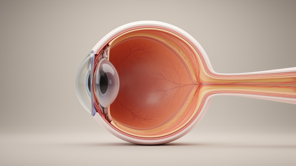
A computational biomechanics study using whole-eye finite element models has revealed that the anatomical subtype of posterior scleral staphyloma exerts the primary influence on mechanical deformation across the lamina cribrosa and macular region in eyes affected by pathologic myopia. Investigators established three-dimensional numerical simulations capturing five classical morphological subtypes alongside variations in axial length and intraocular pressure. Across the evaluated parameters, the specific geometry of the staphyloma subtype demonstrated a substantially greater statistical effect on structural deformation than axial elongation alone. These findings demonstrate that localized outpouching configurations alter intraocular pressure-driven stress distributions, offering a biomechanical explanation for why clinical complications and visual prognoses diverge markedly among patients presenting with comparable degrees of overall eyeball elongation.
Pathologic myopia represents a leading cause of irreversible visual impairment worldwide, driven by excessive axial elongation of the eyeball that induces severe vision-threatening complications including myopic maculopathy, choroidal neovascularization, retinal detachment, and glaucoma. Posterior scleral staphyloma constitutes a hallmark structural abnormality of the disease, defined by an abnormal outpouching of the posterior ocular wall caused by progressive scleral thinning and tissue ectasia. This localized wall bulging inflicts mechanical deformation on crucial posterior pole structures, particularly the neural retina, choroid, fovea, and optic nerve head. Topographically, staphylomas exhibit considerable structural heterogeneity, historically categorized into distinct subtypes according to the classification established by Curtin. Although clinicians have long recognized correlative associations between staphyloma classifications and clinical fundus damage, the precise underlying biomechanical mechanisms and localized strain distributions have remained largely unaddressed.
To investigate these mechanical interactions under controlled physical conditions, the research team constructed whole-eye three-dimensional finite element models incorporating nine distinct anatomical ocular components: the cornea, sclera, ciliary body, choroid, retina, lamina cribrosa, optic nerve, crystalline lens, and zonules. The baseline models adopted a uniform transverse diameter of twenty-five millimeters, while scleral, choroidal, and retinal thicknesses were assigned regional values reflecting documented anatomical thinning within the staphyloma, outside the staphyloma, across the optic nerve head, and at the fovea. Scleral thickness measured half a millimeter outside the outpouching but thinned to fifteen hundredths of a millimeter within the staphyloma and at the optic nerve head. Lamina cribrosa thickness was standardized to one hundred micrometers across all models. Mechanical simulations incorporated five primary Curtin-defined staphyloma subtypes: Type I (wide macular), Type II (narrow macular), Type III (peripapillary or optic nerve head), Type IV (nasal), and Type V (inferior).
The computational matrix incorporated three distinct axial lengths—twenty-six millimeters, twenty-nine millimeters, and thirty-six millimeters—representing mild, moderate, and extreme axial elongation commonly documented in pathologic myopia cohorts. Morphological staphyloma variations were parameterized using the physical coordinates of the bulging origin, along with specific outward depth and surface radius dimensions tailored to each subtype. Linear isotropic material properties were assigned to most ocular tissues, including an elastic modulus of three megapascals for the sclera, six tenths of a megapascal for the choroid, three hundredths of a megapascal for the retina and optic nerve, and three tenths of a megapascal for the lamina cribrosa. Boundary conditions restrained the corneoscleral junction while establishing a frictionless contact interface between the lateral optic nerve and scleral canal. Whole-eye models were subsequently solved under intraocular pressure loading states of ten and twenty-one millimeters of mercury using ANSYS Mechanical software, generating a dataset of three hundred and forty static simulations.
To capture regional biomechanical insults while minimizing numerical discretization errors associated with extreme point stresses, the researchers evaluated four primary response metrics. Optic nerve head deformation was assessed via the mean displacement in the radial direction of the lamina cribrosa and the mean displacement in the circumferential direction. Macular insult was evaluated using the high-strain ratio, defined as the percentage of elements within a seventy-five hundredths millimeter radius of the macular center experiencing an equivalent strain equal to or exceeding four percent, alongside the internal strain ratio measuring core foveal strain contribution. Global analysis of the model cohort demonstrated overall mean radial lamina cribrosa displacement of 12.21 ± 9.26 micrometers and mean circumferential displacement of 9.88 ± 9.50 micrometers. Peak equivalent strains systematically localized along the anatomical interface between the neural retina and the lamina cribrosa.
Non-parametric statistical evaluations utilizing the Kruskal–Wallis test revealed that staphyloma subtype-specific geometry accounted for the largest proportion of mechanical response variability across the tested parameter ranges, displaying effect sizes between 0.335 and 0.619. In sharp contrast, axial length yielded effect sizes below 0.136, demonstrating that eyeball elongation alone plays a secondary role relative to regional staphyloma contouring. Significant intergroup differences emerged among the five subtypes across all key metrics. Type I wide macular staphylomas, which encompass both the macular center and the optic nerve head, generated the largest lamina cribrosa deformations, exhibiting mean radial displacements of 0.0215 ± 0.0133 millimeters and circumferential displacements of 0.0188 ± 0.0138 millimeters. Conversely, Type V inferior staphylomas exhibited the lowest lamina cribrosa deformations, averaging 0.0078 ± 0.0035 millimeters radially and 0.0057 ± 0.0037 millimeters circumferentially.
Receiver operating characteristic analyses demonstrated robust discriminative performance of these biomechanical metrics across simulated conditions. For distinguishing an elevated intraocular pressure state of twenty-one millimeters of mercury from ten millimeters of mercury, peak optic nerve head equivalent strain yielded an area under the curve of 0.993, while the macular high-strain ratio achieved an area under the curve of 0.883. Furthermore, when classifying Type I staphyloma against all other configurations, radial lamina cribrosa displacement demonstrated an area under the curve of 0.811 with a sensitivity of 64.1 percent and a specificity of 88.41 percent. In differentiating macular-involving subtypes (Types I and II) from non-macular subtypes, the high-strain ratio separated groups with an area under the curve of 0.831, reflecting lower overall baseline strain exposure within the broad, less abruptly curved macular staphyloma basins compared to peripapillary configurations.
Analysis of staphyloma depth and radius dimensions revealed that structural contouring governs localized tissue stretching in a subtype-specific manner. Following permutation testing and multivariate linear regression across non-surgical models, staphyloma radius emerged as the dominant geometrical driver, showing strong positive correlations with radial displacement and macular high-strain ratios, with Spearman coefficients ranging from 0.74 to 0.95.
The study also quantitatively evaluated the mechanical impact of posterior scleral reinforcement, a surgical intervention aimed at halting staphyloma progression through the placement of an exogenous supporting graft. Simulating a single five-millimeter-wide, one-millimeter-thick reinforcing strip applied symmetrically across the macular center at twenty-six millimeters of axial length, the researchers varied the strip elastic modulus across eight levels ranging from three to fifty megapascals. Surgical intervention substantially reduced intraocular pressure-driven macular high-strain exposure across all five staphyloma subtypes, dropping overall mean high-strain ratios from 19.45 percent down to 3.58 percent, corresponding to relative reductions between 23.5 percent and 87.7 percent. Higher strip moduli produced monotonic decreases in macular strain accumulation. However, macular strip placement produced no statistically significant reduction in radial or circumferential lamina cribrosa displacements, indicating that macular reinforcement primarily stabilizes the fovea without relieving anterior-posterior or canalicular optic nerve traction.
The authors noted several methodological simplifications inherent to their static idealized modeling framework. The lamina cribrosa was modeled with a uniform thickness of one hundred micrometers without incorporating microarchitectural pore geometry or physiological tilting. Additionally, ocular scleral tissue was represented as a homogeneous, linear, isotropic material, omitting the complex collagen fiber alignments, non-linear tensile stiffening, and equatorial anisotropy characteristic of native human sclera. Furthermore, the simulated surgical models assessed static reinforcement configurations at a single axial length without accounting for postoperative dynamic eye movements or biological integration.
Subject of Research: Finite element biomechanical modeling of posterior scleral staphyloma subtypes and posterior scleral reinforcement in pathologic myopia
Article Title: Biomechanical responses and scleral reinforcement across staphyloma subtypes in pathologic myopia: a finite element analysis
Article References: Wang, Y., Li, X., Ren, J., Qiao, L., & Qian, X. (2026). Biomechanical responses and scleral reinforcement across staphyloma subtypes in pathologic myopia: a finite element analysis. Frontiers in Bioengineering and Biotechnology, 14, Article 1948781. https://doi.org/10.3389/fbioe.2026.1948781
Image Credits: AI Generated
DOI: 10.3389/fbioe.2026.1948781
Keywords: pathologic myopia, posterior scleral staphyloma, finite element analysis, posterior scleral reinforcement, lamina cribrosa displacement, macular strain, biomechanics, Curtin classification, optic nerve head, intraocular pressure, scleral modulus, ocular elongation, ophthalmology simulation, myopic maculopathy, computational ophthalmology, axial length
News Source: Gregory Coleman. (November 14, 2026). Eye Models Reveal Staphyloma Subtypes Drive Ocular Strain in Pathologic Myopia. Scienmag.