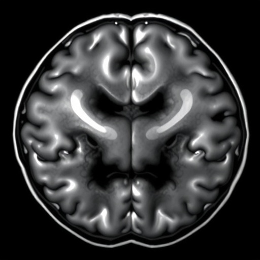
In the ever-evolving landscape of medical imaging, the need for precise and efficient segmentation of brain tumors from MRI scans has never been more critical. With the prevalence of various types of brain tumors, early diagnosis and effective treatment planning hinge on high-quality imaging modalities. A recent survey conducted by Yang et al. sheds light on U-Net variant networks, which have garnered attention for their striking effectiveness in segmenting brain tumors from MRI scans. This exploration highlights not just advancements in technology but also the collaborative efforts that are pushing the boundaries of medical research.
The U-Net architecture, initially designed for biomedical image segmentation, has emerged as a cornerstone in various medical imaging applications. Its unique structure—a contracting path that captures context and a symmetric expanding path that enables precise localization—has made it particularly adept at handling the complexities of MRI scans. The architecture facilitates multi-scale feature extraction, which is essential when dealing with the diverse presentations of brain tumors in patients. As Yang and his colleagues delve into the various adaptations of this architecture, it becomes apparent that understanding these modifications is crucial for future advancements in the field.
One of the most striking aspects of U-Net’s performance lies in its ability to outperform traditional segmentation methods, often achieving higher accuracy and better localization capabilities. By leveraging convolutional neural networks (CNN), U-Net variants effectively capture intricate features that may be overlooked by less sophisticated algorithms. This deep learning approach enables the models to analyze vast quantities of image data, facilitating the extraction of meaningful patterns that inform clinical decision-making processes. The proficiency of these neural networks is particularly relevant as medical imaging becomes increasingly reliant on AI and machine learning technologies.
Moreover, the modifications made to the original U-Net architecture are noteworthy. Researchers have proposed numerous enhancements, including the integration of residual connections, attention mechanisms, and multi-scale feature extraction techniques. These innovations allow the U-Net variants to adapt to the unique characteristics of brain tumors, which often present with varying shapes and sizes. As a result, they enhance the model’s robustness, providing reliable segmentation outputs that can significantly impact patient outcomes.
Furthermore, the survey underscores the importance of extensive training datasets in improving the performance of U-Net variants. High-quality, annotated datasets are indispensable for training deep learning models effectively; they ground the algorithms in reality, allowing them to learn from diverse examples. As more datasets become publicly available, researchers can better train and validate their models, pushing the efficacy of U-Net applications to new heights. This democratization of data is a crucial factor in fostering collaboration among research institutions, ultimately enhancing the quality of outputs and the reliability of findings.
In analyzing the various U-Net adaptations, the survey highlights the role of transfer learning, where models pretrained on large datasets can be fine-tuned to specialize in brain tumor segmentation. This strategy not only speeds up the training process but also helps in mitigating the challenges posed by limited available data. The ability to leverage knowledge gained from related tasks endows researchers with a powerful tool, driving better performance in niche applications like brain tumor imaging.
As we grapple with the complexities of brain tumor management, the implications of these technological advancements are profound. Accurate segmentation can inform treatment planning, guide surgical interventions, and even assist in monitoring tumor progression or regression throughout a patient’s treatment journey. The insights gleaned from Yang et al.’s survey illuminate a path toward harnessing the full potential of U-Net variants in clinical settings, enhancing the ability to provide timely and effective interventions for patients.
Emerging evidence suggests that the integration of U-Net variants into clinical workflows may lead to a paradigm shift in how we approach brain tumor diagnostics and treatment. Radiologists could leverage AI-driven segmentation tools to complement their assessments, ensuring that critical information is not missed. This could reduce the cognitive load on medical professionals, allowing them to focus on more complex decision-making processes while the algorithm efficiently handles image segmentation tasks.
However, it is also essential to recognize the challenges that accompany the deployment of such technologies in clinical practice. Issues surrounding model interpretability, validation across diverse populations, and compliance with regulatory standards must be addressed to ensure the safe adoption of AI-driven segmentation tools. Yang et al.’s work serves as a reminder that while the technology holds immense promise, careful consideration of ethical and practical implications must guide its deployment.
As the conversation around U-Net variants continues to evolve, collaboration among researchers, clinicians, and technology developers will be key to unlocking their full potential. Symposiums and workshops dedicated to AI in medical imaging can foster an environment conducive to innovation, allowing for the cross-pollination of ideas that can drive the field forward. Encouraging interdisciplinary collaboration is vital in tackling the multi-faceted challenges posed by brain tumor diagnosis and treatment.
In conclusion, Yang et al. provide a compelling overview of U-Net variants in the context of MRI brain tumor segmentation. Their findings pave the way for further exploration and innovation in the realm of medical imaging. As we look to the future, the potential of these technologies to transform patient care is considerable. The journey toward realizing this potential will require persistence, collaboration, and a commitment to ethical standards, but the rewards could be monumental in the quest for improved outcomes in brain tumor management.
The implications of this research are profound, as it highlights a shift toward a future where AI not only assists in diagnosing conditions but also enhances the quality of life for patients battling brain tumors. The integration of sophisticated deep learning models into everyday clinical practice could soon become a reality, bridging the gap between technological advancement and patient care. The future is indeed bright for the intersection of artificial intelligence and medical imaging, as innovations continue to unfold.
In sum, the survey conducted by Yang et al. acts as a pivotal reference for both researchers and practitioners in understanding the potential of U-Net variants in MRI brain tumor segmentation. This comprehensive overview underscores the value of advancing our methodologies through the lens of modern technology, ultimately contributing to improved patient outcomes.
Subject of Research: MRI Brain Tumor Segmentation Using U-Net Variants
Article Title: A survey of U-Net variant network for MRI brain tumor segmentation
Article References:
Yang, W., Zhang, R., Chow, S.K.K. et al. A survey of U-Net variant network for MRI brain tumor segmentation.
Discov Artif Intell (2025). https://doi.org/10.1007/s44163-025-00525-0
Image Credits: AI Generated
DOI:
Keywords: MRI, Brain Tumor, U-Net, Segmentation, Artificial Intelligence, Deep Learning, Medical Imaging, Neural Networks
Tags: advancements in MRI technologybiomedical image segmentation techniquesbrain tumor diagnosis and treatmentcollaborative efforts in medical researchearly diagnosis of brain tumorseffective imaging modalities for brain tumorsinnovations in brain tumor analysismachine learning in medical imagingMRI brain tumor segmentationmulti-scale feature extraction in medical imagingU-Net architecture for medical imagingU-Net variants for image segmentation
