Recent advancements in biomedical engineering have introduced significant innovations in the understanding and modeling of human anatomy, particularly concerning the head and neck region. At the forefront of this field, a recent study by Zhou, Reddy, and Yin stands out for its intricate exploration of the head–neck musculoskeletal model. This research integrates the complex dynamics of cervical spine rhythms, offering unprecedented insights measured through in vivo dynamic radiography.
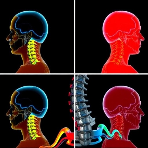
The cervical spine, a critical structure supporting the head, is not only responsible for mobility but also plays a pivotal role in maintaining neural and vascular integrity. Traditionally, studies of cervical motion have relied on static images and generalized models that lacked the precision needed for detailed understanding. The new model proposed by the authors incorporates dynamic radiographic measurements, highlighting the significance of real-time data in comprehending the multifaceted movements of the cervical region.
Dynamic radiography presents numerous advantages over conventional imaging techniques. By capturing motion in real time, it allows for a detailed assessment of the cervical spine’s biomechanics during various activities. This method is particularly transformative as it enables researchers to observe the spine in action, evaluating how different forces and motions influence its structure and function. The integration of this technology into the musculoskeletal model offers a level of detail that enhances the accuracy of biomechanical analyses.
One of the key innovations in this research is the improved musculoskeletal model that better reflects the complexities of human anatomy. By integrating dynamic measurements, the authors have crafted a model that accounts for the rhythmic motions of the cervical spine, which are essential during everyday activities like turning the head or nodding. This nuanced approach allows for a more accurate depiction of how the cervical spine interacts with surrounding musculature and tissues, thereby paving the way for more effective treatments for neck injuries or disorders.
Moreover, the study delves into the implications of these findings on clinical practices. With a deeper understanding of cervical spine dynamics, healthcare professionals can develop better diagnostic tools and therapeutic strategies tailored to individual patients’ needs. This could lead to more personalized rehabilitation programs, enhancing recovery outcomes for those suffering from neck pain or injuries resulting from trauma or degeneration.
An important aspect of the research is its dedication to understanding how cervical rhythms influence not only mobility but also the overall quality of life. Chronic neck pain, often stemming from poor cervical spine biomechanics, can significantly impact daily living. By providing a clearer picture of the underlying mechanics, the authors offer new pathways to address such issues, potentially alleviating pain and improving the functional capabilities of patients.
Beyond its immediate implications for clinical practice, this research opens avenues for deeper investigations into other areas of human biomechanics. The methodologies developed could be applied to other regions of the musculoskeletal system, further enriching our understanding of human motion. The potential cross-applicability of these findings could ultimately lead to comprehensive models that encompass the entire body, offering insights into how various systems interact during movement.
Furthermore, the innovative combination of advanced imaging techniques and sophisticated biomechanical modeling underscores the importance of interdisciplinary collaboration in biomedical research. This study exemplifies how engineers, medical professionals, and researchers can come together to address complex biological questions, demonstrating the power of collective expertise in furthering healthcare solutions.
In an era where personalized medicine is gaining traction, the ability to model and simulate individual variations becomes paramount. The head–neck musculoskeletal model presented in this research serves as a prototype for future advancements in personalized healthcare. By considering the unique anatomical and biomechanical characteristics of each patient, practitioners may provide more effective, individualized care strategies that take into account the nuanced ways in which different patients’ bodies function.
As the research community continues to explore the intricacies of human biomechanics, the emphasis on real-time, dynamic data acquisition will likely gain momentum. Future studies building on these findings may incorporate machine learning algorithms to predict outcomes based on the newly created models, providing healthcare professionals with powerful tools for diagnosis and treatment planning.
In summary, Zhou and colleagues’ study represents a pivotal advancement in our understanding of the head and neck musculoskeletal system. By integrating dynamic radiographic measurements into their model, they have enhanced our grasp of cervical spine mechanics, with broad implications for both clinical applications and further research. This innovative approach has the potential to revolutionize how we understand and treat cervical spine disorders, ultimately improving patient care across a multitude of healthcare settings.
As this field of study matures, the integration of technology and biomechanical models will pave the way for exciting new developments. The ongoing challenge will be to translate these insights into practice, ensuring that patients benefit from the latest scientific advancements. As highlighted by this research, the future of biomedical engineering is bright, filled with possibilities that extend beyond the current limitations of our understanding.
The commitment to advancing knowledge in the field of biomechanics, evidenced by this research, reflects a growing recognition of the importance of evolving medical technologies. As new methodologies continue to emerge, one can anticipate a shift toward increasingly precise, dynamic models that will shape the future of patient care in ways previously thought unattainable.
This innovative study is not just a testament to the technological capabilities of modern science but also a reminder of the incredible complexities of the human body. Understanding these complexities, and translating that understanding into tangible healthcare improvements, can lead to a paradigm shift in how we approach musculoskeletal health across all demographics.
With the publication of this research, the authors have set a new standard for studies involving dynamic models of the cervical spine, inspiring future explorations that will undoubtedly benefit from their foundational work. The ongoing dialogue among researchers, clinicians, and educators in this field is critical as we strive to unravel the complexities of human motion and develop ever more sophisticated approaches to medical science.
Subject of Research: Head–Neck Musculoskeletal Model Incorporating Cervical Spine Rhythms
Article Title: An Improved Head–Neck Musculoskeletal Model Incorporating Cervical Spine Rhythms Measured by Dynamic Radiography In Vivo
Article References: Zhou, Y., Reddy, C., Yin, W. et al. An Improved Head–Neck Musculoskeletal Model Incorporating Cervical Spine Rhythms Measured by Dynamic Radiography In Vivo. Ann Biomed Eng (2026). https://doi.org/10.1007/s10439-026-03971-8
Image Credits: AI Generated
DOI: https://doi.org/10.1007/s10439-026-03971-8
Keywords: Cervical Spine, Musculoskeletal Model, Dynamic Radiography, Biomechanics, Patient Care
Tags: advancements in cervical spine researchbiomechanics of cervical spinecervical spine dynamicsdetailed assessment of cervical motiondynamic radiography advantageshead neck musculoskeletal modelin vivo dynamic measurementsinnovations in biomedical engineeringmusculoskeletal modeling in anatomyneural and vascular integrity of cervical spinereal-time imaging techniquesunderstanding head neck region


