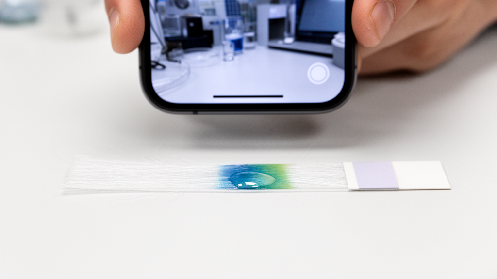
A research team has developed an instrument-free colorimetric spot-test platform based on functionalized polymer nanofibers that allows for the rapid preliminary screening of hydrogen peroxide in coastal seawater. Fabricated by blending poly-ε-caprolactone with poly(ethylene glycol) diglycidyl ether through electrospinning, the resulting composite mats provide a robust, highly hydrophilic substrate that immobilizes horseradish peroxidase in a single operational step without requiring additional chemical activating agents. When exposed to microliter volumes of sample alongside a chromogenic co-substrate, the dry membrane produces an unmistakable green coloration whose intensity correlates with peroxide concentration. By capturing the resulting spot color with an everyday mobile phone camera and processing the image data through standard analytical software, the researchers established a linear quantitative detection range spanning from 0.05 to 1.0 millimolar hydrogen peroxide, achieving a calculated limit of detection of 0.004 millimolar. This design demonstrates how combining accessible electrospun nanomaterials with enzyme-driven color reactions can eliminate the need for delicate benchtop optical systems during initial on-site water quality assessments.
Hydrogen peroxide represents the most abundant and comparatively long-lived reactive oxygen species found in marine environments, functioning as an essential indicator of photochemical activity, organic matter turnover, and environmental stress. In sunlit coastal surface waters, the molecule is generated primarily through the photochemical oxidation of chromophoric dissolved organic matter and direct biological release by marine organisms, while being systematically broken down through natural enzymatic decay and catalytic pathways. Consequently, ambient marine concentrations reflect a dynamic balance between local photochemical processes, microbial turnover, and hydrodynamic mixing. Tracking this compound provides vital data for monitoring water quality, evaluating the transformation of organic pollutants, and charting the redox cycling of trace metals. However, established quantitative techniques such as high-performance chromatography, fluorometry, and benchtop spectrophotometry present severe logistical hurdles for fieldwork. These conventional laboratory protocols demand bulky instrumentation, light-sensitive derivatization reagents, stable optical benches, trained technicians, and extensive sample pretreatment to mitigate saline matrix interference, all while risking sample composition changes during storage and transit from coastal or shipboard collection sites.
To overcome these field limitations, the study leveraged electrospinning to create a self-supporting, high-surface-area fibrous support capable of stabilizing enzymes and confining tiny reagent volumes to uniform analytical spots. Poly-ε-caprolactone was chosen as the base structural material due to its proven mechanical durability, biodegradability, and dependable fiber-forming characteristics during electrospinning. Nevertheless, pristine electrospun poly-ε-caprolactone exhibits pronounced hydrophobicity, displaying static water contact angles typically between 114 and 129 degrees. This extreme water repellency prevents aqueous droplets from wetting the membrane, thereby obstructing dissolved target molecules from reaching immobilized biological catalysts. To resolve this surface barrier, the researchers blended poly(ethylene glycol) diglycidyl ether directly into the pre-spinning solution. The biocompatible poly(ethylene glycol) backbone of this secondary modifier inherently imparts strong hydrophilicity to the final blend, while its twin terminal epoxide functional groups offer accessible covalent anchoring sites for protein attachment throughout the three-dimensional fiber network.
Fabrication of the fibrous matrix involved systematically evaluating precursor mixtures to optimize structural uniformity and avoid structural collapse. Poly-ε-caprolactone was dissolved at a fixed concentration of 10 percent weight-to-volume in a binary solvent system containing formic acid and acetone in a 3 to 7 volume ratio. Specific quantities of poly(ethylene glycol) diglycidyl ether were subsequently added to yield molar concentrations of 0.071, 0.177, and 0.354 molar before the mixtures were stirred overnight at 37 degrees Celsius and ultrasonicated. Electrospinning was conducted at an applied voltage of 10 kilovolts, a constant solution flow rate of 1.0 milliliter per hour, and a collector distance of approximately 12 centimeters under controlled environmental conditions of 22 to 23 degrees Celsius and 58 to 65 percent relative humidity. Scanning electron microscopy revealed that while a lower concentration of 0.071 molar yielded a broad, bimodal fiber diameter distribution between 0.10 and 0.38 micrometers, the 0.177 molar composition produced smooth, continuous, bead-free fibers with diameters ranging from 0.2 to 0.75 micrometers. Conversely, increasing the modifier content to 0.354 molar caused excessive fiber fusion and loss of distinct fibrous boundaries, cementing 0.177 molar as the optimal formulation.
The physical transformation of the electrospun material was corroborated through surface wettability and water absorption experiments. While native poly-ε-caprolactone fibers strongly resist water uptake with a baseline swelling ratio of only 0.03, the engineered composite with 0.177 molar poly(ethylene glycol) diglycidyl ether exhibited a dramatic water contact angle reduction down to 10.885 ± 0.28 degrees, demonstrating near-complete surface wetting. In continuous liquid immersion tests over 26 hours, the composite mats achieved an equilibrium swelling ratio of roughly 1000 to 1100 percent within the first hour of contact, absorbing approximately ten times their dry mass in water without losing structural integrity or disintegrating. Fourier-transform infrared spectroscopy confirmed the integration of both polymer constituents into the finished fibers, showing characteristic ester carbonyl stretching at 1723 reciprocal centimeters alongside ether backbone vibrations at 1097 reciprocal centimeters. This hydrophilic architecture ensures that applied sample droplets are drawn instantaneously into the porous fibrous mesh, keeping the enzyme, co-substrate, and target analyte confined in close physical contact during testing.
Enzyme functionalization was achieved using a single-step coupling procedure that avoids secondary chemical crosslinkers or surface-activating reagents. Horseradish peroxidase and bovine serum albumin were prepared separately in 50 millimolar sodium acetate buffer at pH 5.5 and combined in an equal volume ratio. A single 1-microliter droplet of this protein mixture was deposited directly onto the composite mat and allowed to dry at room temperature for approximately three minutes, creating a ready-to-use test zone without subsequent washing. In this formulation, bovine serum albumin serves an essential structural function rather than acting as an inert filler. Its accessible amine groups react with a portion of the exposed epoxide rings on the fiber surfaces, consuming reactive sites and preventing the excessive multipoint covalent attachment of horseradish peroxidase that often causes protein rigidification and loss of catalytic turnover. Furthermore, the added albumin elevates the overall protein content of the 1-microliter droplet, enabling it to spread evenly across the hydrophilic mat and dry into a compact, repeatable spot with well-defined boundaries rather than forming an irregular, diffuse ring.
Surface-sensitive analytical techniques confirmed that the catalytic proteins were successfully retained on the electrospun fibers. Scanning electron microscopy verified that while the open porous network remained intact following protein deposition, the fiber surfaces took on a denser, coated appearance indicative of a dry proteinaceous overlayer. X-ray photoelectron spectroscopy survey scans demonstrated the appearance of distinctive nitrogen 1s, sulfur 2p, and iron 2p core-level signals over the polymer background, which was matched by energy-dispersive X-ray spectroscopy measurements recording 5.43 weight percent nitrogen, 1.01 weight percent sulfur, and 0.14 weight percent iron. Because neither pristine poly-ε-caprolactone nor poly(ethylene glycol) diglycidyl ether contains these heteroatoms, their presence confirms the surface protein layer. While nitrogen and sulfur originate from peptide bonds and amino acid residues common to both albumin and horseradish peroxidase, the 0.14 weight percent iron signature is exclusively diagnostic of the single iron protoporphyrin IX heme group located within the peroxidase active site, proving that active enzyme molecules were securely integrated onto the test mat.
The colorimetric assay relies on the catalytic reduction of hydrogen peroxide coupled to the oxidation of 2,2-azino-bis(3-ethylbenzothiazoline-6-sulfonic acid) diammonium salt, which acts as a reducing co-substrate. Under optimized buffer conditions at pH 5.5, the catalytic cycle initiates with a two-electron oxidation of the resting ferric enzyme by hydrogen peroxide, producing water and generating the high-valent oxoferryl porphyrin cation radical intermediate designated as Compound I. This intermediate subsequently undergoes two sequential one-electron reduction steps by abstracting electrons from two separate molecules of the colourless substrate dianion, passing through a Compound II intermediate and yielding two molecules of the intensely green radical anion alongside a second water molecule. For analytical readout, 1 microliter of 3 millimolar co-substrate solution in pH 7.4 phosphate buffer and 1 microliter of sample solution were spotted sequentially onto the dry enzyme spot. Once the chromogenic reaction occurred, spots were photographed under uniform light-emitting diode ring illumination inside a controlled black imaging box at a fixed distance of 12 centimeters using an iPhone 15 smartphone, followed by circular region-of-interest mean color intensity analysis within ImageJ.
Systematic calibration showed that the color development exhibited distinct qualitative and quantitative stages across increasing analyte concentrations. Visual inspection distinguished three clear response classes: spots treated with 0.05 millimolar or less hydrogen peroxide remained entirely colourless, spots receiving 0.4 to 0.6 millimolar turned light green, and those exposed to 1.0 millimolar developed an intense dark green hue. Quantitative evaluation of the green color channel within a standardized circular area yielded a linear calibration relationship described by the equation y = 807.6x + 1033.1 with a coefficient of determination of 0.9317 across the range of 0.05 to 1.0 millimolar. Above 1.0 millimolar, the optical signal reached a plateau up to 2.0 millimolar due to enzyme active-site saturation, followed by a slight decrease in color intensity at 3.0 millimolar. This high-concentration suppression aligns with the well-known reversible inactivation of horseradish peroxidase, in which excess peroxide drives the formation of the catalytically dormant oxyperoxidase state known as Compound III.
To verify that complex natural waters would not disrupt the enzymatic spot test, the researchers evaluated real-world applicability using natural coastal seawater collected directly from İzmir Bay at Karşıyaka and from Seferihisar Bay, running parallel comparative assays on local municipal tap water and commercial bottled drinking water. Because typical baseline concentrations of hydrogen peroxide in coastal surface waters fall within nanomolar to micromolar ranges, the trial was structured as a matrix tolerance spike-recovery challenge using an added peroxide concentration of 0.5 millimolar in undiluted samples. The resulting analytical recoveries ranged cleanly between 94 and 104 percent across all tested matrices, demonstrating that the high ionic strength, dissolved salts, and background organic matter present in raw marine water do not cause significant matrix interference or suppress color development on the functionalized membrane.
Long-term practical utility for distributed testing hinges on how well immobilized biomolecules retain their biocatalytic function during storage. The researchers tracked the operational longevity of the enzyme-loaded nanofiber mats by storing prepared strips under dry conditions at 4 degrees Celsius and testing their colorimetric response against a fixed peroxide concentration over a 10-day period. Signal retention was calculated relative to freshly prepared mats evaluated on day zero. Over the full 10-day testing window, the mats exhibited only a minor 5.3 percent loss in total colorimetric activity, demonstrating that the covalent entrapment network provided by the epoxide-functionalized poly(ethylene glycol) chains and bovine serum albumin framework effectively shields horseradish peroxidase from conformational degradation during refrigerated storage.
Despite these promising capabilities, the authors noted several clear analytical boundaries defining the current scope of the technology. Most notably, because the linear operational range spans 0.05 to 1.0 millimolar with a detection limit of 0.004 millimolar, the current system cannot directly detect the nanomolar background concentrations typical of pristine open oceans without a dedicated preconcentration step or more sensitive optical transduction. Furthermore, while the presence of epoxide functional groups provides a sound chemical rationale for single-step covalent bonding, the interfacial chemical bonds were not directly differentiated from physical adsorption through specialized surface binding spectroscopy. Finally, minor spot-to-spot variations arising from liquid droplet drying dynamics on the fibrous texture contribute to the observed scatter in calibration measurements. Nevertheless, as a low-cost, portable screening device requiring only a smartphone camera, the platform provides an effective tool for tracking elevated peroxide levels in industrial discharge, coastal oxidation events, and marine stress monitoring.
Subject of Research: Colorimetric detection of marine hydrogen peroxide using enzyme-functionalized electrospun nanofibers and smartphone imaging
Article Title: Peroxidase-immobilized polycaprolactone/poly(ethylene glycol) diglycidyl ether electrospun nanofibers for colorimetric detection of hydrogen peroxide in seawater
Article References: Kilic Kolbasi, N. M., & Odaci, D. (2026). Peroxidase-immobilized polycaprolactone/poly(ethylene glycol) diglycidyl ether electrospun nanofibers for colorimetric detection of hydrogen peroxide in seawater. Discover Chemistry, 3(1), Article 637. https://doi.org/10.1007/s44371-026-01089-3
Image Credits: AI Generated
DOI: 10.1007/s44371-026-01089-3
Keywords: hydrogen peroxide, colorimetric detection, electrospun nanofibers, horseradish peroxidase, polycaprolactone, polyethylene glycol diglycidyl ether, seawater analysis, spot test, smartphone sensing, marine biogeochemistry, reactive oxygen species, ABTS, bovine serum albumin, swelling ratio, water contact angle, spike recovery, surface wettability, enzyme immobilization
News Source: Bethany Barker. (November 18, 2026). Engineered Nanofiber Spot Test Detects Marine Hydrogen Peroxide Using Smartphone Imaging. Scienmag.