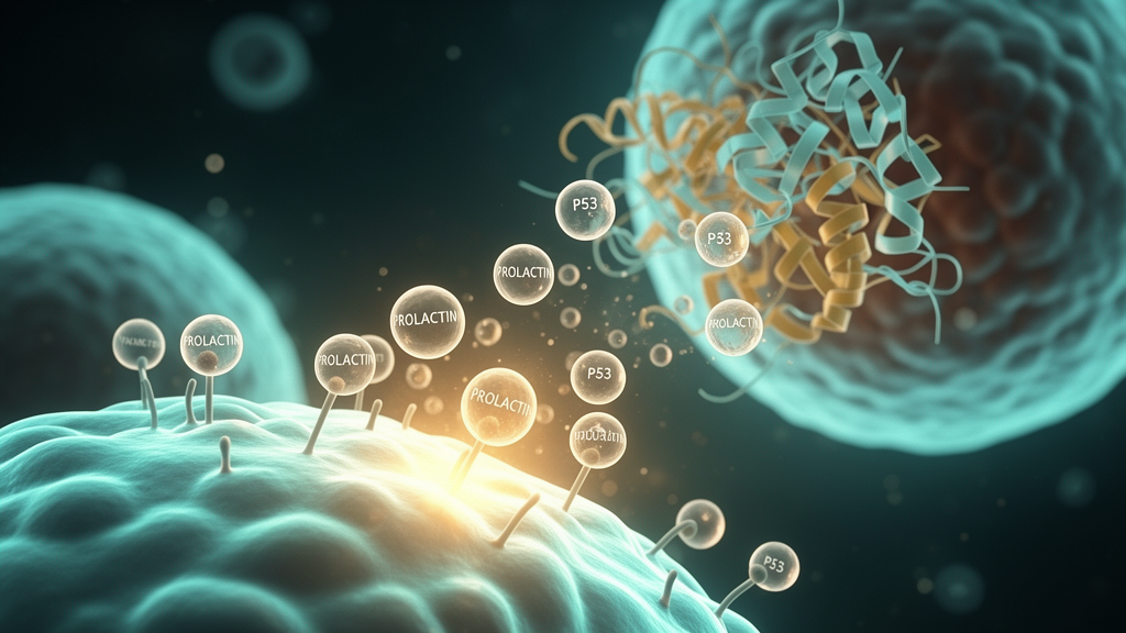
A multicenter case-control study conducted across tertiary care hospitals in Kerala has identified significant elevations in serum prolactin concentrations and p53 gene expression among individuals experiencing assisted reproductive technology failure compared with fertile controls. The clinical investigation, encompassing 600 adult participants, demonstrated that while both molecular and endocrine markers were substantially altered in cases of repeated reproductive failure, their relationship is characterized by a weak positive association marked by considerable inter-individual variability rather than a direct, p53-driven causal mechanism. Serum prolactin emerged as a robust predictive biomarker of reproductive outcome, displaying particularly strong discriminatory accuracy among female participants.
The study cohort was recruited between 2022 and 2024 from the departments of reproductive medicine and assisted reproductive technology units alongside general health and postnatal outpatient clinics. The investigation enrolled 300 infertile individuals comprising 150 couples with a mean age of 34.5 ± 4.9 years, alongside 300 fertile controls comprising 150 couples with a mean age of 34.5 ± 4.6 years. Infertility was clinically defined according to World Health Organization criteria as the inability to achieve a clinical pregnancy following at least twelve months of regular unprotected intercourse. Cases were specifically categorized as assisted reproductive technology failure following the absence of clinical pregnancy across at least two completed in vitro fertilization or intracytoplasmic sperm injection cycles involving the transfer of morphologically good-quality embryos. Controls were frequency-matched by age and sex in a one-to-one ratio, with confirmed natural fertility established by having at least one naturally conceived child and no prior history of infertility or assisted reproductive interventions.
To isolate relevant biological signals and limit potential confounding influences, the research team established rigorous participant selection criteria. Individuals presenting with confirmed genetic disorders, active malignancies, chronic systemic illnesses, or primary endocrine and pituitary pathologies—including documented thyroid dysfunction—were excluded from enrollment. The exclusion protocol also disqualified candidates undergoing current hormonal therapies or taking dopamine agonists, psychotropic medications, or any pharmacological agents known to alter endogenous prolactin or gonadotropin secretion at the time of sampling. These criteria ensured that measured hormonal variations reflected underlying reproductive pathology rather than secondary pharmacological or systemic endocrine disruption.
Standardized biological sampling procedures were implemented to account for physiological fluctuations and circadian rhythms. Fasting venous blood samples measuring 8 to 10 mL were drawn exclusively between 8:00 AM and 10:00 AM to minimize diurnal variation in circulating prolactin levels. For all female participants, venipuncture was restricted to the early follicular phase, specifically between menstrual cycle days two and five. Furthermore, blood collection among assisted reproductive technology cases was conducted strictly prior to the commencement of exogenous ovarian stimulation protocols, preventing gonadotropin medications from masking baseline endocrine and molecular profiles.
Laboratory evaluation combined targeted quantitative gene expression assays with dual-antibody hormonal quantification. Total ribonucleic acid was isolated from peripheral blood samples using a commercial guanidine thiocyanate–phenol spin-column extraction kit, with sample purity verified by A260/A280 spectrophotometric ratios between 1.8 and 2.0. Complementary deoxyribonucleic acid synthesis was conducted using 50 μg of total RNA primed with oligo(dT)18 and random hexamers. Quantitative real-time polymerase chain reaction was executed with SYBR Green master mix across thirty-two amplification cycles, adhering to MIQE guidelines with primer amplification efficiencies maintained between 95% and 105%. Relative p53 gene expression was normalized against the glyceraldehyde-3-phosphate dehydrogenase endogenous control gene utilizing the comparative 2^(-ΔΔCt) calculation method. In parallel, circulating concentrations of prolactin, follicle-stimulating hormone, and luteinizing hormone were measured in duplicate using sandwich enzyme-linked immunosorbent assays pre-coated with human hormone-specific antibodies.
The study design was statistically powered based on a standard sample size formulation accounting for an estimated general infertility prevalence of 10% and a 3% margin of error at a 95% confidence level, securing an 80% statistical power threshold. Analytical evaluations were executed in Stata version 17.0 and RStudio, utilizing independent sample t-tests supplemented with bootstrapping techniques to calculate group differences, 95% confidence intervals, and Cohen’s d effect sizes. Given the fundamental physiological dimorphism governing mammalian endocrinology, all statistical assessments and subsequent receiver operating characteristic curve evaluations were conducted independently for male and female cohorts, with statistical significance established at p < 0.001 across all primary markers.
Among female participants, comparative analyses revealed substantial hormonal and transcriptional shifts between cases and controls. Serum prolactin exhibited the most pronounced elevation among infertile women, yielding a test statistic of t = 18.43, a 95% confidence interval spanning 15.62 to 21.24, and a very large effect size of Cohen’s d = 2.135. Peripheral p53 gene expression was similarly upregulated in female cases (t = 9.47, 95% CI: 7.76 to 11.17, Cohen’s d = 1.097). Circulating luteinizing hormone showed significant elevation in cases compared with controls (t = 7.73, 95% CI: 5.15 to 10.31, Cohen’s d = 0.895), while follicle-stimulating hormone exhibited a moderate group difference (t = 4.51, 95% CI: 2.19 to 6.84, Cohen’s d = 0.523), confirming widespread endocrine and transcriptional alteration in women experiencing repeated implantation or cycle failure.
Parallel assessments within the male cohort confirmed corresponding, highly significant differences across all evaluated parameters. Prolactin concentrations were markedly increased in male cases relative to fertile controls, demonstrating a 95% confidence interval of 9.23 to 12.78 and a large effect size of Cohen’s d = 1.275. Relative p53 gene expression was elevated in infertile men with a 95% confidence interval of 6.80 to 10.00 and an effect size of d = 0.974. Furthermore, male cases displayed elevated gonadotropins, with luteinizing hormone presenting a 95% confidence interval of 6.44 to 9.41 (Cohen’s d = 0.918) and follicle-stimulating hormone demonstrating a 95% confidence interval of 5.65 to 9.16 (Cohen’s d = 0.858), confirming systemic endocrine shifts across both partners in affected couples.
Receiver operating characteristic curve analyses demonstrated that circulating prolactin possesses exceptional diagnostic discrimination for identifying assisted reproductive technology failure, particularly in women. In the female cohort, prolactin achieved an area under the curve of 0.9181, operating with 89% sensitivity and 95% specificity, accompanied by elevated positive likelihood ratios and suppressed negative likelihood ratios. Female p53 gene expression demonstrated robust predictive value with an area under the curve of 0.8128 (75% sensitivity, 81% specificity), outperforming luteinizing hormone (AUC: 0.7738, 81% sensitivity, 77% specificity) and follicle-stimulating hormone (AUC: 0.6666). In males, prolactin delivered an area under the curve of 0.828 at a clinical cut-off concentration of 20.38 ng/mL (69% sensitivity, 81% specificity), while male p53 expression yielded an area under the curve of 0.787 (67% sensitivity, 89% specificity), surpassing male luteinizing hormone (AUC: 0.733) and follicle-stimulating hormone (AUC: 0.726).
Despite marked elevations across both parameters, regression and scatter plot evaluations uncovered only a weak positive correlation between prolactin concentrations and p53 gene expression among clinical cases. The data distribution within the case group displayed substantial inter-individual scattering, whereas fertile controls exhibited tightly clustered data points with low baseline p53 expression that remained stable across varying prolactin levels. Similarly weak associations were observed between p53 expression and circulating gonadotropins, including luteinizing hormone and follicle-stimulating hormone, in both sexes. Female cases exhibited higher transcript variability than controls across mid-range hormone concentrations, pointing to complex biological heterogeneity rather than a direct, uniform regulatory relationship.
These analytical findings directly challenge hypotheses postulating direct transcriptional or causal regulation between p53 and prolactin secretion in reproductive failure. The authors note that while previous literature has documented p53 pathway involvement in embryonic implantation, apoptosis, and pituitary adenomas, the weak correlation identified in peripheral blood samples indicates that p53 does not function as an immediate upstream regulator of circulating prolactin. Instead, the concurrent elevation of these biomarkers is more consistent with parallel endocrine and molecular dysregulation, potentially arising from shared cellular stress responses, upstream regulatory disruptions, or multi-pathway endocrine abnormalities.
The investigators emphasized important methodological boundaries, classifying the study as hypothesis-generating due to its observational, expression-based design. Because peripheral gene expression does not establish mechanistic causality, direct functional validation within reproductive tissues remains necessary before clinical translation. Furthermore, recruitment was confined to tertiary medical centers within a single geographic region in Kerala, and unmeasured residual confounding cannot be entirely dismissed in multifactorial reproductive disorders. Future research incorporating multi-center longitudinal cohorts, diversified patient demographics, and functional laboratory models will be required to determine whether these concurrent biomarkers can inform individualized risk stratification and clinical monitoring protocols in assisted reproduction.
Subject of Research: Association of p53 gene expression and reproductive hormones with assisted reproductive technology failure
Article Title: Molecular insights into p53-driven hormonal dysregulation and ART failure
Article References: Nair, R. K. R., Josephine, A., Sureka, V., Varghese, A. C., Pillai, S. P., Veluthakunju, A. E., Chettiyar, A. D., & Divakaran, D. R. (2026). Molecular insights into p53-driven hormonal dysregulation and ART failure. Egyptian Journal of Medical Human Genetics, 27(1), Article 41. https://doi.org/10.1186/s43042-026-00874-0
Image Credits: AI Generated
DOI: 10.1186/s43042-026-00874-0
Keywords: Assisted Reproductive Technology, Infertility, Prolactin, p53 Gene Expression, In Vitro Fertilization, Intracytoplasmic Sperm Injection, Follicle-Stimulating Hormone, Luteinizing Hormone, Biomarkers, Case-Control Study, Endocrine Dysregulation, Kerala, Receiver Operating Characteristic, Real-Time PCR, Enzyme-Linked Immunosorbent Assay, Implantation Failure, TP53, Hyperprolactinemia
News Source: Juliet Wilcox. (November 13, 2026). Elevated Prolactin and p53 Expression Are Associated With Assisted Reproductive Technology Failure. Scienmag.