Conducted at a level I trauma center, the clinical investigation tracked patients who underwent simultaneous medial closing-wedge distal femoral osteotomy and medial closing-wedge high tibial osteotomy to treat complex, bifocal valgus malalignments between 2011 and 2015. Group-level radiographic evaluations revealed that the mean mechanical tibiofemoral angle shifted substantially from a pronounced preoperative valgus deformity of 9.6 ± 2.6 degrees down to −1.4 ± 1.5 degrees at six weeks following surgical correction, settling at a neutral mean of 0.1 ± 4.7 degrees at a mean mid-term follow-up of 7.5 ± 1.2 years.
Morphological examinations have confirmed that substantial valgus malalignment is frequently bifocal in nature, stemming from simultaneous structural deformities situated in both the distal femur and the proximal tibia.
The study protocol evaluated a clinical series of 12 patients representing 13 operative knees who underwent varization double-level osteotomy, with a mandatory minimum follow-up duration established at five years. Over the longitudinal evaluation period, five patients accounting for five operative knees were unavailable for final mid-term reassessment, with four patients unable to be contacted and one patient having died, resulting in an overall patient follow-up rate of 58.3 percent. The final evaluated mid-term cohort comprised seven patients with eight treated knees examined after a mean postoperative follow-up duration of 7.5 ± 1.2 years, spanning an overall range from 5.7 to 9.4 years. In the single patient who underwent bilateral surgical corrections, an interval of at least one full year was strictly enforced between the two operative procedures to guarantee complete physical recovery and independent functional rehabilitation. The clinical investigation was conducted under formal approval granted by the local ethics committee, with all participating subjects providing written informed consent prior to study enrollment.
The clinical team utilized a mechanical medial proximal tibial angle greater than 90 degrees and a mechanical lateral distal femoral angle below 85 degrees as morphological benchmarks indicating tibial and femoral deformity contributions, although these values were applied as clinical guides rather than mandatory isolated cutoffs. Preoperative digital simulations were systematically performed to model single-level distal femoral or high tibial osteotomies; when achieving the target mechanical axis of neutral to 2 degrees of varus alignment would induce a non-physiological periarticular angle, double-level osteotomy was selected.
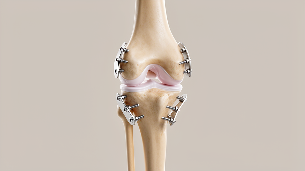
The surgical intervention commenced with diagnostic knee arthroscopy to directly inspect intra-articular cartilage surfaces, confirm the primary surgical indication, and exclude undetected intra-articular pathologies. Following arthroscopy, distal femoral correction was executed via a minimally invasive biplanar medial closing-wedge distal femoral osteotomy accessed through a 5 to 6 centimeter medial incision using a subvastus approach with blunt preparation toward the medial cortex. The calculated medial bone wedge, averaging 5.4 ± 2.0 mm in planned height, was excised, and the osteotomy was closed around the lateral osseous hinge before rigid internal fixation with an angular-stable TomoFix medial distal femur plate.
The tibial osteotomy was completed in a biplanar fashion, executing two precise oblique cuts toward the fibular head accompanied by an ascending cut positioned directly behind the patellar tendon. After removing the predetermined medial bone wedge, which possessed a planned mean height of 6.3 ± 1.2 mm, the osteotomy was closed around the intact lateral hinge and stabilized using an angular-stable TomoFix medial high tibia plate equipped with locking screws. Intraoperative mechanical alignment and joint-line orientation were verified using fluoroscopy and a long alignment rod prior to layered wound closure. Postoperative rehabilitation enforced partial weight-bearing of 20 kilograms starting on day one, advancing to full weight-bearing after approximately six weeks once radiographic consolidation was verified, avoiding external orthoses in favor of active physical therapy.
Detailed radiographic assessments confirmed marked coronal corrections at both the group level and across individual joint parameters. The mean preoperative mechanical tibiofemoral angle of 9.6 ± 2.6 degrees valgus was corrected to −1.4 ± 1.5 degrees at six weeks postoperatively and measured 0.1 ± 4.7 degrees at the final mid-term examination. Preoperative femoral deformity, represented by a mean mechanical lateral distal femoral angle of 86.1 ± 2.2 degrees, improved to 90.5 ± 2.5 degrees at six weeks and remained stable at 90.2 ± 2.4 degrees at final follow-up. Similarly, the mean mechanical medial proximal tibial angle shifted from an abnormal baseline of 92.4 ± 1.6 degrees down to 86.4 ± 1.5 degrees postoperatively, measuring 87.3 ± 2.1 degrees at mid-term evaluation. Despite consistent group-level stability, individual alignment trajectories exhibited measurable heterogeneity. In knee 5, the planned mechanical axis of −0.5 degrees deviated to −4.1 degrees postoperatively, paired with an achieved femoral angle of 95.8 degrees against a planned 92.0 degrees. Furthermore, knee 8 demonstrated recurrent valgus alignment reaching 9.8 degrees at final follow-up, accompanied by an increase in the joint-line convergence angle from 3.0 to 9.1 degrees and a change in mMPTA from 85.8 to 90.3 degrees.
However, subsequent hardware removal was performed across all eight evaluated knees after an average postoperative interval of 3.2 ± 2.9 years, spanning a range of 0.3 to 7.6 years, which the authors emphasized represents an important secondary surgical burden requiring clear preoperative patient counseling.
Functional evaluation and clinical examination at final follow-up revealed no pathological signs of joint effusion, ligamentous laxity, soft-tissue compromise, or restricted range of motion among the evaluated knees. Subjective functional questionnaires reflected moderate residual symptoms alongside high overall treatment contentment, with the Lysholm score reaching 82.5 ± 12.8 points, the Oxford Knee Score averaging 36.2 ± 9.9 points, and the International Knee Documentation Committee subjective score achieving 65.2 ± 25.4 percent. The comprehensive Knee Injury and Osteoarthritis Outcome Score totaled 322.1 ± 129.3 points, with subscale scores of 69.1 ± 24.6 for pain, 73.2 ± 20.6 for symptoms, 74.4 ± 23.6 for activities of daily living, 47.5 ± 42.2 for sports and recreation, and 57.8 ± 26.0 for quality of life. When surveyed regarding their overall satisfaction, all patients stated they would choose to undergo the double-level procedure again, with 37.5 percent describing themselves as satisfied, 62.5 percent as delighted, 75.0 percent reporting met expectations, and 25.0 percent reporting exceeded expectations.
The study authors highlighted several methodological limitations that temper the clinical interpretation and generalizability of their mid-term findings. The retrospective observational design lacked a comparative control group undergoing single-level osteotomy or primary joint arthroplasty, and functional scores were collected solely at final follow-up, preventing longitudinal statistical comparisons of patient-reported functional recovery over time. A critical limitation involves the small sample size and substantial loss to follow-up, where only 58.3 percent of originally treated patients completed mid-term radiographic and functional reviews. Comparative baseline analysis revealed that patients lost to follow-up were younger on average at 41.4 ± 8.1 years compared to 46.5 ± 13.7 years for followed patients, but had higher body mass index values averaging 37.6 ± 6.6 kg/m² versus 31.1 ± 3.4 kg/m², leaving the direction and magnitude of potential selection bias unresolved. Additionally, Kellgren–Lawrence radiographic grading was conducted without blinding to patient identity or examination timeline, yielding substantial intrarater reliability with a Cohen’s kappa of 0.737 but only moderate interrater reliability of 0.539, underscoring the exploratory nature of the findings and the need for larger prospective comparative trials.
Subject of Research: Mid-term radiographic and functional outcomes of varization double-level osteotomy for severe valgus knee deformity
Article Title: Maintenance of periarticular joint angles after varization double-level osteotomy for severe valgus deformity: a technical and radiographic mid-term case series
Article References: Herbst, M., Schröter, S., Ateschrang, A., Histing, T., & Ahrend, M.-D. (2026). Maintenance of periarticular joint angles after varization double-level osteotomy for severe valgus deformity: a technical and radiographic mid-term case series. Archives of Orthopaedic and Trauma Surgery, 146(1), Article 403. https://doi.org/10.1007/s00402-026-06539-2
Image Credits: AI Generated
DOI: 10.1007/s00402-026-06539-2
Keywords: varization double-level osteotomy, valgus knee deformity, distal femoral osteotomy, high tibial osteotomy, mechanical tibiofemoral angle, mechanical lateral distal femoral angle, mechanical medial proximal tibial angle, joint-line convergence angle, joint-line obliquity, knee osteoarthritis, orthopaedic surgery, TomoFix plate, Kellgren-Lawrence grading, Lysholm score, Oxford Knee Score, KOOS, IKDC score
News Source: Ophelia Keating. (November 12, 2026). Double-Level Osteotomy Maintains Periarticular Knee Alignment in Severe Valgus Deformity. Scienmag.