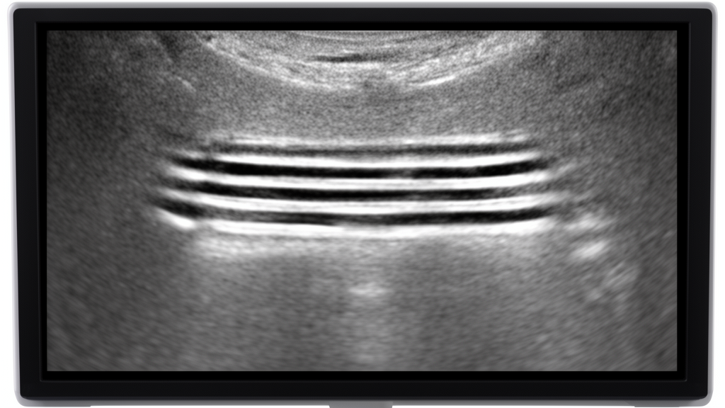
Parallel bands appearing in regurgitant Doppler spectra, known as tiger stripes, are a common and benign artifact rather than a sign of serious intracardiac pathology, according to a retrospective study published in Echo Research & Practice. The research, conducted by Biswaranjan Mishra, analyzed 4,567 consecutive transthoracic echocardiograms (TTEs) performed on adults between 18 and 65 years of age at a single center. The study found that these spectral patterns occurred in 5.4% of the studies, with a 95% confidence interval of 4.8% to 6.1%. This finding challenges the traditional clinical interpretation that tiger stripes indicate oscillating structures such as vegetations or flail leaflets, which have historically prompted urgent further evaluation for potentially grave conditions.
The study was designed to determine the frequency, clinical importance, and possible mechanism of tiger stripes in a routine, unselected echocardiography population. Prior reports had largely described these findings in highly selected patients with prosthetic valve dysfunction or discrete oscillating lesions, leading to an assumption that the stripes were markers of significant pathology. However, systematic data on the frequency and relevance of tiger stripes in a broad, consecutive population remained limited. The authors aimed to clarify whether these spectral patterns were associated with specific valvular pathologies or if they represented a reproducible physical artifact unrelated to structural abnormalities.
The retrospective review included TTE studies performed between January 23, 2024, and January 24, 2025, using a GE Vivid 8 platform. Inclusion criteria required adult patients aged 18 to 65, with images analyzed for the presence of tiger stripes in both pulsed-wave (PW) and continuous-wave (CW) Doppler spectra of valvular flows. Exclusion criteria included inadequate Doppler quality, congenital lesions requiring non-standard windows, and intracardiac devices causing uninterpretable artifacts. The definition of a tiger stripe was specific: the presence of at least two parallel, echo-dense bands superimposed on a regurgitant Doppler spectrum, at approximately equal spacing, on one or both sides of the baseline, visible in at least two consecutive cardiac cycles.
The primary reviewer analyzed all studies blinded to clinical data beyond the valve label. To ensure reliability, a limited interobserver validation study was conducted with two qualified cardiologists who independently reviewed a subset of 100 transthoracic echocardiograms, comprising 50 cases with tiger stripes and 50 without. The interobserver agreement for tiger stripe detection was 91%, with a Cohen’s kappa coefficient of 0.82 (95% CI, 0.73–0.91), indicating almost perfect agreement. This high level of consistency supports the reproducibility of the artifact identification process in clinical settings.
The results showed that tiger stripes occurred predominantly in left-sided regurgitant lesions, specifically mitral regurgitation (MR) and aortic regurgitation (AR), where regurgitant spectral velocity is high. Notably, stripes were not detected in non-hypertensive TR, pulmonary regurgitation, stenotic lesions, or diastolic flow across atrioventricular valves. The study also found that stripes were more conspicuous in mild to moderate jets and higher-velocity Doppler spectra, suggesting a relationship between flow dynamics and the visibility of the artifact.
A critical finding of the study was the absence of any association between tiger stripes and oscillating intracardiac structures. None of the cases with tiger stripes were associated with vegetations, flail leaflets, or other oscillating masses in the regurgitation pathway. Conversely, tiger stripes were not noted in patients where oscillating structures were present, such as in cases of vegetation, ruptured chordae, or prosthetic valve dehiscence. This inverse relationship contradicts earlier hypotheses that linked the stripes to the motion of these structures, indicating that the artifact is likely generated by flow dynamics rather than tissue oscillation.
The authors proposed a hypothetical mechanism involving the formation of red blood cell (RBC) rouleaux during flow convergence proximal to the regurgitant orifice. Slowing of blood flow can lead to the aggregation of RBCs into rouleaux, which act as strong reflectors of ultrasound waves. These parallel aggregates may generate reverberation and mirror-image artifacts, creating the characteristic parallel bands. The high-pitched cooing or whistling sound often associated with tiger stripes may result from returning high-frequency sound waves interacting with these aggregates. However, the authors emphasize that this mechanism remains speculative and was not directly tested in the present study.
The study also examined the audible Doppler signals associated with tiger stripes. All cases with tiger stripes were associated with a high-pitched cooing or whistling sound in the returning audible signal. However, there was no association with clinical musical murmurs on auscultation. This distinction is important because prior literature had sometimes correlated the audible Doppler signal with musical murmurs caused by vibrating intracardiac structures. The lack of correlation with clinical murmurs further supports the idea that tiger stripes are a spectral artifact rather than a manifestation of structural vibration.
The study has several limitations, including its single-center, retrospective design, which may introduce selection bias. The use of a single imaging platform and a limited interobserver sample size also restricts the generalizability of the findings. Additionally, the proposed mechanism for the artifact is hypothetical and requires further investigation. The authors did not perform a formal systematic review of the literature, meaning that some prior reports on tiger stripes may not have been captured.
The clinical implications of these findings are significant for echocardiographers and cardiologists. Recognizing tiger stripes as a common artifact can help avoid unnecessary investigations for presumed oscillating masses when no corroborating imaging signs exist. The presence of tiger stripes should not be used to grade the severity of regurgitation or to infer the mechanism of valvular disease. This understanding aligns with recent guidelines on the assessment of prosthetic valve dysfunction, which do not mention tiger stripes as an indication of valve malfunction.
The study contributes to the broader understanding of Doppler artifacts in cardiac imaging. Artifacts such as reverberation, acoustic shadowing, and mirror-image phenomena are well-documented, but tiger stripes have often been misinterpreted as pathological signs. By providing a large-scale analysis of this specific artifact, the study clarifies its frequency and clinical irrelevance.
The research highlights the importance of distinguishing between true pathological findings and imaging artifacts in clinical practice.
In conclusion, tiger stripes in Doppler echocardiography are a common, benign spectral artifact that should not prompt additional imaging for oscillating masses or be used to grade regurgitation severity. This finding has important implications for clinical practice, helping to prevent unnecessary investigations and misdiagnoses. Further research is needed to fully elucidate the physical mechanisms behind these artifacts, but the current evidence strongly supports their benign nature in routine echocardiography.
Subject of Research: Frequency and clinical significance of tiger stripe artifacts in transthoracic echocardiography
Article Title: Tiger stripe: an innocuous Doppler artifact
Article References: Mishra, B. (2026). Tiger stripe: an innocuous Doppler artifact. Echo Research & Practice, 13(1), Article 2. https://doi.org/10.1186/s44156-026-00104-8
Image Credits: AI Generated
DOI: 10.1186/s44156-026-00104-8
Keywords: Doppler echocardiography, Tiger stripes, Cardiac imaging, Doppler artifact, Mitral regurgitation, Aortic regurgitation, Clinical cardiology, Ultrasound physics, Tiger, stripe, innocuous, Doppler
News Source: Ophelia Keating. (November 9, 2026). Doppler tiger stripes are common artifacts, not pathology. Scienmag.