A recent clinical case report published in Discover Immunity documents the successful diagnosis and management of secondary hemophagocytic lymphohistiocytosis triggered by disseminated histoplasmosis in a 57-year-old immunocompromised man. The patient, who had a medical history of rheumatoid arthritis managed with infliximab, methotrexate, and prednisone, along with osteoporosis and depression, initially presented to an outside hospital experiencing fevers, chills, and myalgias. Upon presentation, his vital signs showed a body temperature of 101 degrees Fahrenheit, a blood pressure of 145/80 mmHg, a heart rate of 110 beats per minute, and a respiratory rate of 18 breaths per minute, while appearing in minimal acute distress. His outpatient immunosuppressive medications were promptly discontinued, and high-dose corticosteroid therapy was initiated after an initial infectious workup yielded negative results.
The patient was readmitted two days later with recurrent fevers and acute liver injury, evidenced by aspartate aminotransferase and alanine aminotransferase levels rising into the 600s. Although initial blood testing indicated Epstein-Barr virus-related hepatitis, a subsequent liver biopsy evaluated with hematoxylin and eosin alongside Grocott’s methenamine silver staining confirmed the presence of histoplasmosis. Additional clinical evidence, including examination of a peripheral blood smear and a positive urine histoplasma antigen assay, supported the diagnosis of disseminated histoplasmosis. Clinicians initiated treatment with oral itraconazole while gradually tapering the patient off corticosteroids, but the fungal infection progressed despite this targeted intervention.
Shortly after beginning itraconazole monotherapy, the patient required hospitalization again for persistent fevers, worsening myalgias, gastrointestinal bleeding, and severe cytopenias. Laboratory monitoring revealed a decline in hemoglobin from 11.7 g/dL to 8.0 g/dL, while his platelet count dropped sharply from 103,000/µL to 16,000/µL. Diagnostic imaging and endoscopies revealed multiple severe systemic abnormalities, including gastritis identified on esophagogastroduodenoscopy, a hepatic subcapsular hematoma, and renal infarction. Computed tomography ruled out a pulmonary embolism in the setting of acute hypoxia, though it demonstrated ground-glass opacities and nodular lung disease, alongside clinically detectable splenomegaly, a ferritin measurement of 5550 ng/mL, and serum triglyceride concentrations reaching into the 1000s.
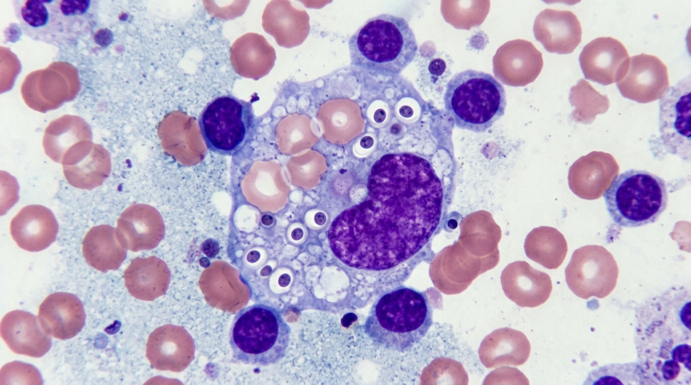
Upon transfer to a specialized medical center, the patient remained febrile, tachycardic, and pancytopenic, raising critical concerns for disseminated intravascular coagulation. After supportive management allowed platelet counts to exceed 50,000/µL, a transesophageal echocardiogram was performed to evaluate for infective endocarditis given the recent renal infarcts, but cardiac imaging and hypercoagulation testing returned negative. An esophagogastroduodenoscopy performed to assess esophageal and gastric mucosa revealed yeast forms within the lamina propria of the stomach consistent with disseminated fungal disease. Bone marrow aspirate and biopsy further confirmed fungal involvement, showing numerous Histoplasma organisms alongside histiocytes actively exhibiting erythrophagocytosis, which established the diagnosis of secondary hemophagocytic lymphohistiocytosis.
Secondary hemophagocytic lymphohistiocytosis is a life-threatening hyperinflammatory syndrome driven by excessive and inappropriate immune activation, especially involving natural killer cells, which triggers widespread macrophage activation in the bone marrow and hemophagocytosis. Diagnostic standards require patients to fulfill at least five out of eight established clinical criteria, comprising persistent fever, splenomegaly, cytopenias affecting at least two hematopoietic cell lineages, hypertriglyceridemia or hypofibrinogenemia, hemophagocytosis on tissue pathology, decreased natural killer cell activity, elevated serum ferritin above 500 ng/mL, or elevated soluble CD25 concentrations exceeding 2400 U/mL. The patient met criteria through persistent fever, splenomegaly, cytopenias, marked hypertriglyceridemia, diagnostic bone marrow pathology, and extreme ferritin elevation, supported by elevated interleukin-2 receptor levels.
Prompt identification of the underlying infectious trigger is crucial for survival, as secondary hemophagocytic lymphohistiocytosis triggered by disseminated histoplasmosis occurs in less than one percent of cases globally, and treatment delays can be fatal. Histoplasma capsulatum resides primarily in soil contaminated with bird or bat droppings, infecting humans through spore inhalation across endemic regions including the Central and Eastern United States, Central and South America, Africa, Asia, and Australia. While many infections remain asymptomatic or mild in immunocompetent hosts, severe disseminated disease occurs in an estimated 1 in 2,000 individuals, presenting substantial diagnostic complexity when secondary hyperinflammatory cascades develop.
Therapeutic management required switching the patient from oral itraconazole to intravenous liposomal amphotericin B combined with systemic prednisone. Shortly after starting the amphotericin B infusion, the patient experienced a severe delayed infusion reaction characterized by fevers, rigors, chills, tachycardia, and profound hypotension that necessitated vasopressor support and urgent transfer to the medical intensive care unit. Clinicians successfully controlled subsequent adverse events by premedicating the patient thirty minutes prior to each scheduled infusion with acetaminophen, diphenhydramine, meperidine, and intravenous fluid boluses, enabling the completion of the necessary antifungal regimen.
Following two weeks of intravenous liposomal amphotericin B and corticosteroid therapy, the patient showed remarkable clinical stabilization and marked biomarker improvements. His hyperferritinemia improved substantially, with serum ferritin dropping from a peak of 14,900 ng/mL down to 3,000 ng/mL, alongside stabilization of hemoglobin concentrations and complete normalization of platelet counts. With his hyperinflammatory state resolved and hemodynamics stabilized, clinicians transitioned the patient to an oral itraconazole regimen planned for a twelve-month duration in accordance with treatment guidelines from the Infectious Diseases Society of America.
The authors contextualized the clinical findings by reviewing broader evidence concerning treatment outcomes and mortality in histoplasmosis-induced hemophagocytic lymphohistiocytosis. In one evaluated review of 11 patients, four of five individuals who received immunosuppressive therapy died (80 percent), compared to two deaths among five patients who did not receive additional immunosuppression (40 percent), although this comparison was underpowered to establish statistical significance. Another case series examining 60 patients found that 81 percent were managed with amphotericin B formulations, reporting an overall inpatient case fatality rate of 31 percent across the cohort and 37 percent among patients living with HIV, alongside variable mortality rates across subgroups receiving steroids, intravenous immunoglobulin, or chemotherapy agents.
The report emphasizes that considerable therapeutic uncertainty persists regarding the optimal integration of immunosuppressants and intravenous immunoglobulin when secondary hemophagocytic lymphohistiocytosis is driven by acute systemic infections. Because published data derive primarily from non-controlled case series and small observational cohorts, no universal protocol exists for managing disseminated histoplasmosis-induced secondary hemophagocytosis in adults. Clinicians must maintain a high index of suspicion in endemic regions, utilize rapid diagnostic tools such as urine histoplasma antigen tests and tissue biopsies, and tailor immunosuppressive and antifungal therapies carefully to the individual patient’s clinical trajectory.
Beyond histoplasmosis, the literature underscores that secondary hemophagocytic lymphohistiocytosis can arise from an extensive range of infectious pathogens across viral, bacterial, and parasitic categories. Associated infections include human immunodeficiency virus, cytomegalovirus, Epstein-Barr virus, other herpes viruses, dengue, influenza, parvovirus B19, and viral hepatitis. Bacterial and parasitic triggers encompass Mycobacterium tuberculosis, rickettsia species, staphylococcal species, Escherichia coli, Leishmania, malaria, and Toxoplasma species. In evaluating viral drivers, the authors note that while Epstein-Barr virus frequently triggers infectious mononucleosis and secondary hyperinflammation, clinical progression toward full hemophagocytic syndrome depends upon specific host factors, including genetic predispositions and immune regulatory mechanisms. Understanding these distinctions is critical for early identification and targeted management of individuals at risk for severe complications.
Establishing an accurate diagnosis requires integrating molecular, serologic, and histological tools to confirm both the hyperinflammatory process and the underlying pathogen. While diagnostic criteria mandate fulfilling five of eight established features, extreme serum ferritin elevation exceeding 3,000 ng/mL serves as a key diagnostic clue. For confirming Histoplasma capsulatum as the trigger, fungal blood cultures represent the gold standard, whereas urine histoplasma antigen tests provide rapid detection with 90 percent sensitivity. Direct tissue examination through bone marrow and lymph node biopsy offers complementary confirmation by revealing fungal organisms alongside macrophages demonstrating hemophagocytosis. Although immunosuppressed patients face heightened susceptibility to severe disseminated disease, the authors caution that fungal-induced hemophagocytic lymphohistiocytosis can also occur in immunocompetent individuals, highlighting the importance of evaluating travel history to endemic regions across the Americas, Africa, Asia, and Australia.
In the evaluated series of 60 patients with histoplasmosis-induced secondary hemophagocytic lymphohistiocytosis, amphotericin B formulations formed the cornerstone of management, administered to 81 percent of the cohort. Treatment regimens varied widely: nine patients received corticosteroids alone, six received intravenous immunoglobulin alone, three received dexamethasone with etoposide, two received etoposide, dexamethasone, and cyclosporine, two received corticosteroids and intravenous immunoglobulin, and one received anakinra and intravenous immunoglobulin. Mortality differed across therapeutic subgroups: patients treated with amphotericin B had an inpatient mortality rate of 25.0 percent, representing 12 deaths among 48 individuals, while patients receiving corticosteroids had a mortality rate of 31 percent, reflecting five deaths among 16 individuals. Among patients receiving intravenous immunoglobulin, mortality reached 62 percent, or five deaths among eight patients. In contrast, one death occurred among five patients treated with steroids and etoposide with or without cyclosporine, all of whom also received amphotericin B, and the single patient receiving anakinra and intravenous immunoglobulin survived.
Interpreting these clinical outcomes requires acknowledging significant investigative constraints, as non-controlled designs and small cohort sizes prevent definitive conclusions regarding optimal therapeutic combinations. The authors emphasize that currently available evidence cannot resolve whether adjunct immunosuppressive therapy or intravenous immunoglobulin provides a clear survival benefit when severe fungal infection drives hyperinflammation. In the smaller review of 11 cases, the observed mortality difference between patients receiving immunosuppression and those managed without it remained statistically underpowered. Consequently, the investigators highlight an urgent need for larger prospective studies to establish standardized treatment protocols for adult secondary hemophagocytic lymphohistiocytosis, particularly concerning the exact dosing, timing, and role of immunomodulatory agents alongside primary antifungal therapy.
Subject of Research: Clinical management of secondary hemophagocytic lymphohistiocytosis induced by disseminated histoplasmosis
Article Title: Secondary hemophagocytic lymphohistiocytosis to disseminated histoplasmosis in the setting of chronic immunosuppression
Article References: Rodriguez, M., & Patel, A. (2025). Secondary hemophagocytic lymphohistiocytosis to disseminated histoplasmosis in the setting of chronic immunosuppression. Discover Immunity, 2(1), Article 6. https://doi.org/10.1007/s44368-025-00014-7
Image Credits: AI Generated
DOI: 10.1007/s44368-025-00014-7
Keywords: hemophagocytic lymphohistiocytosis, disseminated histoplasmosis, Histoplasma capsulatum, immunosuppression, liposomal amphotericin B, itraconazole, rheumatoid arthritis, hyperferritinemia, Secondary, hemophagocytic, lymphohistiocytosis, disseminated
News Source: Ophelia Keating. (November 6, 2026). Disseminated Histoplasmosis Triggers Rare Hemophagocytic Lymphohistiocytosis in an Immunosuppressed Patient. Scienmag.