In groundbreaking new research published in PLOS Biology, scientists have unveiled compelling evidence that the brain’s isolated cortex, following a surgical procedure called hemispherotomy, exhibits persistent sleep-like slow-wave patterns even during wakefulness. This study, led by Marcello Massimini of Universita degli Studi di Milano along with a multinational team of collaborators, challenges long-held assumptions about brain function and consciousness in disconnected neural tissue.
Hemispherotomy, typically performed on pediatric epilepsy patients with intractable seizures, involves severing communication pathways between one cerebral hemisphere and the rest of the brain. Crucially, the disconnected hemisphere’s cortex remains physically intact with an intact blood supply but is cut off from sensory input and motor output. This surgical disconnection is intended to halt the spread of seizures but also isolates the affected hemisphere, making it behaviorally inaccessible. Until now, the intrinsic activity of this isolated cortex during wakefulness remained a profound mystery.
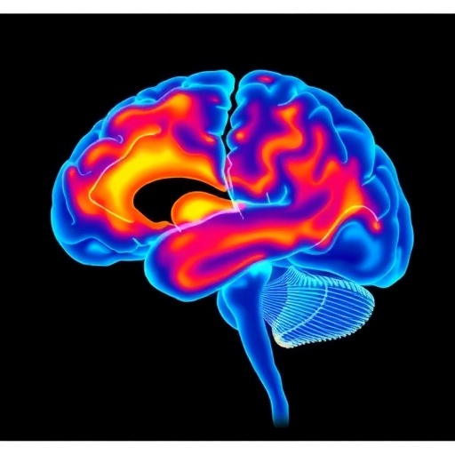
Using scalp electroencephalography (EEG), the researchers analyzed brain activity from ten children who underwent hemispherotomy, spanning periods from immediately before surgery to as long as three years post-operation. Surprisingly, the disconnected hemisphere exhibited pronounced slow-wave activity—characteristic of deep non-rapid eye movement (NREM) sleep—despite the patients being awake. This slow-wave pattern endured over months and years, indicating a lasting alteration in cortical state rather than a transient postoperative phenomenon.
Slow-wave EEG patterns are typically associated with unconscious or poorly conscious states such as deep sleep, general anesthesia, or vegetative states. Their presence in awake patients’ isolated cortex may suggest an absence or severe impairment of internal awareness in the disconnected tissue. Nevertheless, the other, intact hemisphere showed standard wakeful EEG signatures with faster rhythms indicative of active cognition and environmental engagement.
This dichotomy between hemispheres—the isolated cortex locked in a slow-wave, sleep-like mode, versus the connected hemisphere sustaining normal awake patterns—offers a striking example of how brain networks rely on integrated subcortical inputs to maintain conscious states. The researchers emphasize that without activating subcortical structures such as the thalamus, the cortex defaults into this sleep-like baseline, aligning with longstanding theories about the neuronal origins of consciousness and wakefulness.
Importantly, these findings contribute to an ongoing debate about whether behaviorally inaccessible brain regions can host any meaningful conscious experience. The persistence of sleep-like slow waves might indicate a form of cortical “shutdown” with little or no experiential content. However, the team urges caution in drawing direct conclusions about subjective awareness solely based on EEG patterns, especially in tissue completely isolated from sensory and motor pathways.
This work also raises intriguing questions about the potential functional or plastic roles that slow-wave activity might play in isolated brain tissue. Does this persistent sleep-like activity merely reflect a passive loss of input, or could it also support local homeostatic or regenerative processes within the cortex? The study opens new avenues for investigating the interplay between cortical dynamics, consciousness, and neuroplasticity in both health and disease.
The study further underscores the importance of intracranial recordings to complement scalp EEG, especially in clinical contexts requiring close postoperative monitoring. Such invasive methods could provide finer spatial resolution and deeper insights into the electrophysiological states of isolated cortical tissue, ultimately enhancing understanding of patient outcomes and guiding rehabilitative strategies.
Aside from its clinical implications, this research holds profound philosophical significance by challenging notions of consciousness as a unitary, holistic brain state. The emergence of a “sleep-like” island within an otherwise fully awake brain adds complexity to models of subjective experience and highlights the brain’s modular organization with distinct functional compartments.
Researchers Michele A. Colombo, Marcello Massimini, and collaborators articulate the delicate balance and complexity involved in assessing consciousness in neural substrates with no overt behavioral output. Their work elegantly bridges clinical neurology, cognitive neuroscience, and philosophical inquiries into the nature of awareness, contributing valuable empirical data to this challenging field.
The research journey was marked by intense interdisciplinary dialogue and exchange, reflecting the complexity of studying consciousness in isolated neural systems. This multicultural, multi-institutional collaboration spans Italy, Australia, Canada, and the United Kingdom, combining expertise in neuroscience, philosophy, and clinical neurology.
In conclusion, this study provides the first long-term EEG evidence that disconnected cortical regions in hemispherotomy patients reside in a persistent, sleep-like slow-wave state despite wakefulness. These findings have significant ramifications for understanding brain function, consciousness, and rehabilitation strategies following major brain surgery. As research advances, it may unveil new insights into the mechanisms governing cortical state transitions and the elusive neural correlates of conscious experience.
Subject of Research: People
Article Title: Hemispherotomy leads to persistent sleep-like slow waves in the isolated cortex of awake humans
News Publication Date: October 16, 2025
Web References: http://dx.doi.org/10.1371/journal.pbio.3003060
References: Colombo MA, Favaro J, Mikulan E, Pigorini A, Zauli FM, Sartori I, et al. (2025) Hemispherotomy leads to persistent sleep-like slow waves in the isolated cortex of awake humans. PLoS Biol 23(10): e3003060.
Image Credits: Michele A. Colombo (CC-BY 4.0)
Keywords: hemispherotomy, epilepsy surgery, slow-wave sleep, EEG, cortical disconnection, consciousness, pediatric neurology, brain plasticity, neural oscillations, isolated cortex
Tags: brain function and consciousnessdisconnected cerebral hemisphereEEG brain activity analysisepilepsy research breakthroughshemispherotomy surgical procedureimpact of hemispherotomy on brainisolated cortex behavior analysisneurological studies on sleepNREM sleep patterns in awake patientspediatric epilepsy treatmentpersistent brain activity post-surgeryslow-wave brain activity


