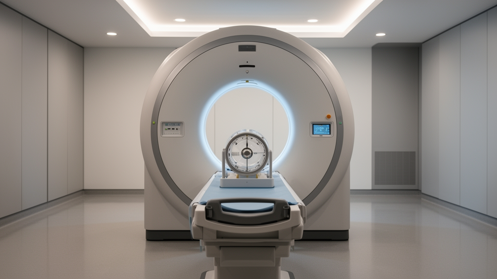
A comprehensive phantom and simulation evaluation has demonstrated that post-treatment Yttrium-90 positron emission tomography on a digital scanner can achieve mean absorbed dose errors below ten percent in large structures, provided that scanner non-linearities are corrected. Conducted on a digital Discovery MI four-ring scanner, the investigation established that quantitative accuracy in selective internal radiation therapy dosimetry relies heavily on matching regularized reconstruction parameters to specific source activity levels. The researchers addressed a central technical obstacle in nuclear medicine oncology, where accurate post-administration radiation mapping is essential to confirm whether radioactive microspheres reached targeted hepatic tumors or inadvertently damaged healthy liver parenchyma. Because the primary hepatic blood supply to tumors originates predominantly from the hepatic artery while normal hepatic tissue is nourished through the portal vein, selective internal radiation therapy utilizes intra-arterially delivered radioisotopes to eradicate malignant lesions. Verifying this targeted delivery through quantitative imaging provides critical clinical confirmation that planned doses match actual delivered radiation distributions.
Assessing Yttrium-90 with positron emission tomography presents distinct physical and technical challenges stemming from the decay properties of the radioisotope itself. Yttrium-90 is primarily a pure beta-minus emitter possessing a physical half-life of 64.2 hours, a maximum beta energy of 2.28 megaelectronvolts, a mean energy of 0.94 megaelectronvolts, and corresponding soft-tissue ranges reaching a maximum of 11 millimeters and a mean of approximately 4 millimeters. Although Yttrium-90 decays almost entirely via beta-minus emission, it features a very rare internal pair production branching ratio that generates positron-electron pairs, producing annihilation photons at 511 kiloelectronvolts that conventional scanners can detect. However, because this branching ratio is exceptionally small, imaging procedures yield very sparse true coincidence counts alongside immense backgrounds of random coincidences and Bremsstrahlung radiation. This background noise is amplified by the intrinsic radioactivity of the scanner lutetium-yttrium oxyorthosilicate crystals. In clinical settings, these low-count statistics lead to severe image noise and pronounced quantitative non-linearities, historically requiring lengthy scanning times on short-axial field-of-view scanners.
To evaluate the quantitative behavior and imaging limitations of digital hardware under clinical conditions, the research team imaged a standardized National Electrical Manufacturers Association International Electrotechnical Commission body phantom across a twelve-day decay sequence. The phantom was initially prepared with 3.64 gigabecquerels of Yttrium-90 citrate, configured with an eight-to-one sphere-to-background activity concentration ratio corresponding to 2.87 megabecquerels per milliliter in the spherical inserts and 0.36 megabecquerels per milliliter throughout the background compartment. Ethylenediaminetetraacetic acid was incorporated into the liquid mixture prior to filling to prevent radioactive adhesion to internal phantom walls. Calibrations for both vials and syringes were conducted using a MEDI-405 dose calibrator traceable to the Laboratoire National Henri Becquerel, with expanded uncertainties of 5.1 percent and 5.5 percent, respectively. Consecutive daily scans tracked activity levels decreasing from 3597 megabecquerels down to 207 megabecquerels at a single bed position, comparing rebinned list-mode acquisition durations of twenty, fifteen, and ten minutes.
Image reconstruction throughout the experimental sequence utilized the proprietary Q.Clear block sequential regularized expectation maximization algorithm, which modulates image smoothing and edge preservation through a single penalty variable designated as the beta parameter. Reconstructions evaluated beta values ranging from 300 to 10,000, incorporating time-of-flight measurements and point spread function modeling while omitting additional post-reconstruction filtering. The PET datasets were reconstructed onto a 256 by 256 matrix with voxel dimensions of 2.73 by 2.73 by 2.79 millimeters within a 700 by 200 millimeter field-of-view, accompanied by standard computed tomography scans at 140 kilovolts peak for attenuation correction. Automated segmentation was executed through an in-house tool utilizing the Insight Toolkit library, establishing theoretical coordinates for each spherical structure, the central lung insert, and 48 sampling spheres positioned throughout the background. Generating unique daily segmentation masks ensured consistent anatomical delineation despite minor day-to-day repositioning of the phantom setup.
Quantitative linearity testing across the twelve-day decay window revealed that measured activity deviated substantially from theoretical activity at both the high and low boundaries of the evaluated range. At activity levels exceeding 2.5 gigabecquerels, the scanner underestimated true phantom activity by 12.9 percent at 2.8 gigabecquerels and up to 15.7 percent at 3.6 gigabecquerels within the phantom volume of interest. Conversely, within an intermediate range between 750 megabecquerels and 2.5 gigabecquerels, quantification remained stable within an acceptable margin of plus or minus ten percent. When activity dropped below 750 megabecquerels, significant overestimation emerged, reaching positive errors of 15.8 percent at approximately 590 megabecquerels and escalating to 52.1 percent at 208 megabecquerels for the phantom volume, while total image volume overestimation reached 88.5 percent. These systematic distortions were tied to random fraction rates of 0.85, 0.90, and 0.94 recorded across high, intermediate, and low activity regimes, where zero-truncation constraints in iterative algorithms convert excessive negative sinogram fluctuations into positive background bias.
To evaluate absorbed dose accuracy independently of scanner artifacts, the authors generated benchmark reference dose maps using Monte Carlo simulations executed via the Geant4 Application for Tomographic Emission version 9.0 platform. The simulation reproduced the exact physical geometry and eight-to-one concentration distribution of the experimental phantom, generating reference dose maps via the Dose Actor at an isotropic voxel resolution of 1.0 millimeter based on eight billion simulated Yttrium-90 decays to keep statistical uncertainty below one percent. These simulations served as the ground truth against which two distinct image-based voxelized dosimetry methods were tested: the Local Deposition Model and the Dose Voxel Kernel approach. The Local Deposition Model assumes complete local kinetic energy absorption within the decay voxel using a derived voxel S-value of 7.181 multiplied by ten to the power of negative nine Gray per Becquerel-second. In contrast, the Dose Voxel Kernel approach convolves activity distributions with a pre-calculated Monte Carlo dose kernel measuring 21 voxels per side.
Protocol optimization was conducted across three representative clinical activity milestones: Day 1 representing a high activity of approximately 3.6 gigabecquerels, Day 6 representing an intermediate activity of approximately 1.0 gigabecquerel, and Day 12 representing a low activity of approximately 200 megabecquerels. Optimal parameter combinations were identified by calculating the Euclidean norm of normalized root mean square errors between image-based and simulated dose-volume histograms for hot spheres and background regions. For high-activity conditions, the analysis demonstrated that acquisitions of fifteen or twenty minutes paired with intermediate beta values between 2000 and 3000 were optimal for the smallest ten-millimeter sphere, whereas larger 37-millimeter structures favored higher beta values between 4000 and 6000. In intermediate-activity settings, twenty-minute acquisitions combined with beta values between 5000 and 7000 for 17-millimeter spheres and 8000 to 10,000 for 37-millimeter spheres yielded superior fidelity. Under low-activity conditions, severe noise required a maximum beta value of 10,000 and a full twenty-minute acquisition to achieve stable convergence.
Evaluating reconstructed spatial resolution via a matched-filter method showed that imaging performance degraded significantly as activity decreased, directly limiting the resolving power achievable in clinical radioembolization. By matching reconstructed sphere recovery coefficients to idealized phantom models filtered with varying Gaussian kernels between 2 and 30 millimeters full width at half maximum, the authors determined effective spatial resolution. Under high-activity conditions, effective spatial resolution measured 7.0 millimeters for protocols optimized on the smallest visible sphere and 10.8 millimeters when optimized on the largest sphere. As activity diminished to intermediate levels, effective spatial resolution widened to 14.8 millimeters and 16.7 millimeters for the respective small- and large-sphere protocols. At the lowest activity tier of 200 megabecquerels, effective spatial resolution degraded to 24.9 millimeters. This substantial widening highlights that count starvation forces regularized algorithms to blur high-frequency details, causing digital PET spatial resolution to approach or fall below conventional single-photon emission computed tomography capabilities under depleted activity conditions.
Direct dosimetric calculations revealed that both voxelized dosimetry techniques systematically underestimated mean absorbed doses across all phantom spheres, driven primarily by partial volume effects and hardware resolution constraints. At high activity, sphere dose underestimation ranged from negative 14.4 percent to negative 69.7 percent, with errors exceeding negative 48.1 percent in spheres seventeen millimeters or smaller when protocols prioritized the largest sphere. Background mean absorbed dose errors remained under 5.5 percent at high activity, but background dose was overestimated by approximately 16 percent under intermediate conditions and by 54 percent at low activity due to non-linearity biases. When images were rescaled to true phantom activity, background dose errors were successfully suppressed to within ten percent across all tiers, dropping below two percent in intermediate regimes, while sphere dose accuracy improved by up to 10.8 percent. Nevertheless, small structures remained heavily impaired by partial volume effects regardless of scaling adjustments.
Comparing the two computational dosimetry approaches indicated that the Local Deposition Model achieved greater dosimetric fidelity in small spherical structures than the Dose Voxel Kernel convolution method. The superior performance of the Local Deposition Model in spheres measuring seventeen millimeters or less aligned with theoretical predictions showing that when system point spread functions exceed the dose kernel dimensions, imaging blur inherently models beta energy transport. In contrast, the Dose Voxel Kernel method exhibited superior dose-volume histogram agreement in large background volumes due to its greater resilience against high-frequency image noise. The authors concluded that while accurate lesion-level dosimetry in small metastases remains constrained by spatial resolution and partial volume loss, implementing model-specific non-linearity corrections enables dependable radiation dosimetry within ten percent error in large hepatic parenchymal and background structures on digital PET scanners.
Subject of Research: Optimization of Yttrium-90 digital PET acquisition and reconstruction parameters for selective internal radiation therapy dosimetry
Article Title: Optimising \(^{90}Y\) PET imaging for dosimetry in SIRT: insights from phantom and simulation studies on the Discovery MI scanner
Article References: Sayah, F., Presles, B., Harb, H., Pham, T.-P., Alberini, J.-L., Popoff, R., & Vrigneaud, J.-M. (2026). Optimising $$^{90}Y$$ PET imaging for dosimetry in SIRT: insights from phantom and simulation studies on the Discovery MI scanner. EJNMMI Physics, 13(1), Article 98. https://doi.org/10.1186/s40658-026-00912-1
Image Credits: AI Generated
DOI: 10.1186/s40658-026-00912-1
Keywords: Yttrium-90, Selective Internal Radiation Therapy, Positron Emission Tomography, Discovery MI, Q.Clear, Local Deposition Model, Dose Voxel Kernel, Monte Carlo Simulation, GATE, Quantitative Linearity, Dose Volume Histogram, NEMA IEC Body Phantom, Spatial Resolution, Partial Volume Effect, Liver Radioembolization, BSREM, Absorbed Dose
News Source: Denise Maddox. (November 12, 2026). Digital PET Dosimetry Achieves Ten Percent Error After Non-Linearity Correction. Scienmag.